
SPEZIELLE VISZERALCHIRURGIE
Facharzt für Viszeralchirurgie und spezielle Viszeralchirurgie
PRECISION. EXPERIENCE. TRUST.
Robotic &
minimally invasive visceral surgery
Specialized minimally invasive visceral surgery combining advanced robotics, extensive operative experience and consistently personal care.
DA VINCI
Fortgeschrittene Erfahrung in der robotergestützten Chirurgie
FORSCHUNG & LEHRE
Habilitation, universitäre Lehre und wissenschaftliche Publikationen
PATIENT IM MITTELPUNKT
Individuelle Beratung und verständliche Behandlungsplanung
15 JAHRE CHIRURGISCHE ERFAHRUNG
Langjährige operative Expertise in der modernen Viszeralchirurgie
LEISTUNGEN & SPEZIALGEBIETE
Spezialisierte Chirurgie. Individuell geplant.
Surgical Specialities
Mein Schwerpunkt liegt auf robotergestützten und minimalinvasiven Verfahren. Jede Behandlung wird anhand der Erkrankung, der anatomischen Situation und der persönlichen Bedürfnisse sorgfältig ausgewählt.
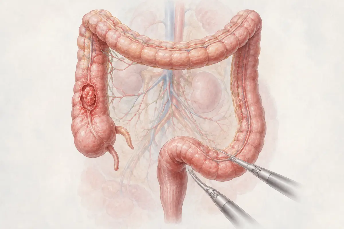
01
Roboterassistierte Kolorektalchirurgie
Robotische Behandlung gutartiger und onkologischer Erkrankungen von Kolon, Rektum und Beckenboden.
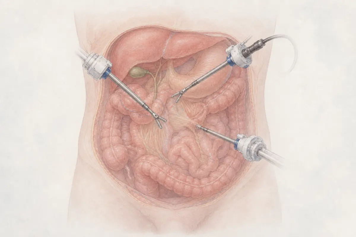
02
Roboterassistierte & minimalinvasive Chirurgie
Moderne chirurgische Verfahren mit Da-Vinci-X- und Xi-Systemen sowie fortgeschrittener Laparoskopie.
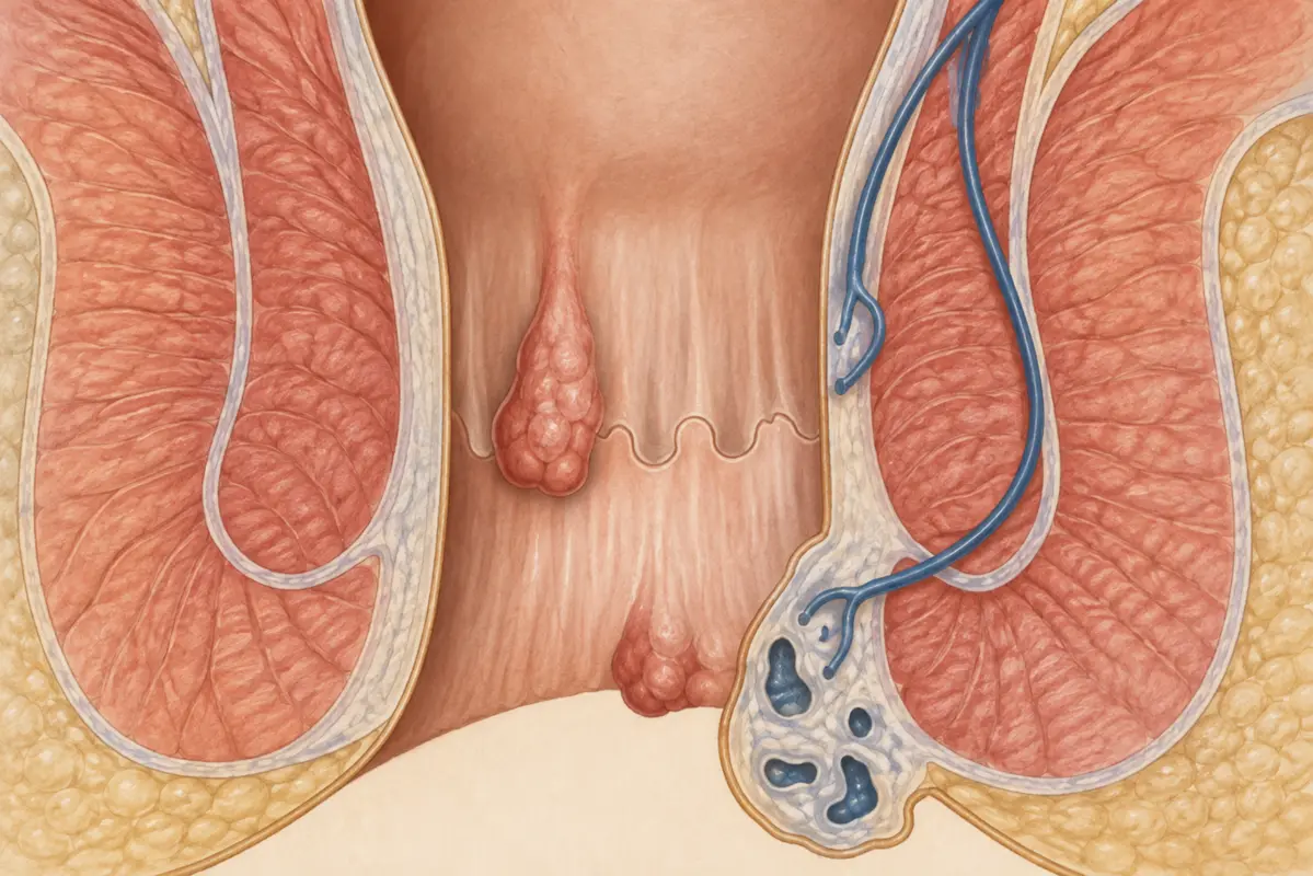
03
Proktologie & Beckenbodenchirurgie
Behandlung funktioneller und struktureller Erkrankungen des Enddarms und Beckenbodens.
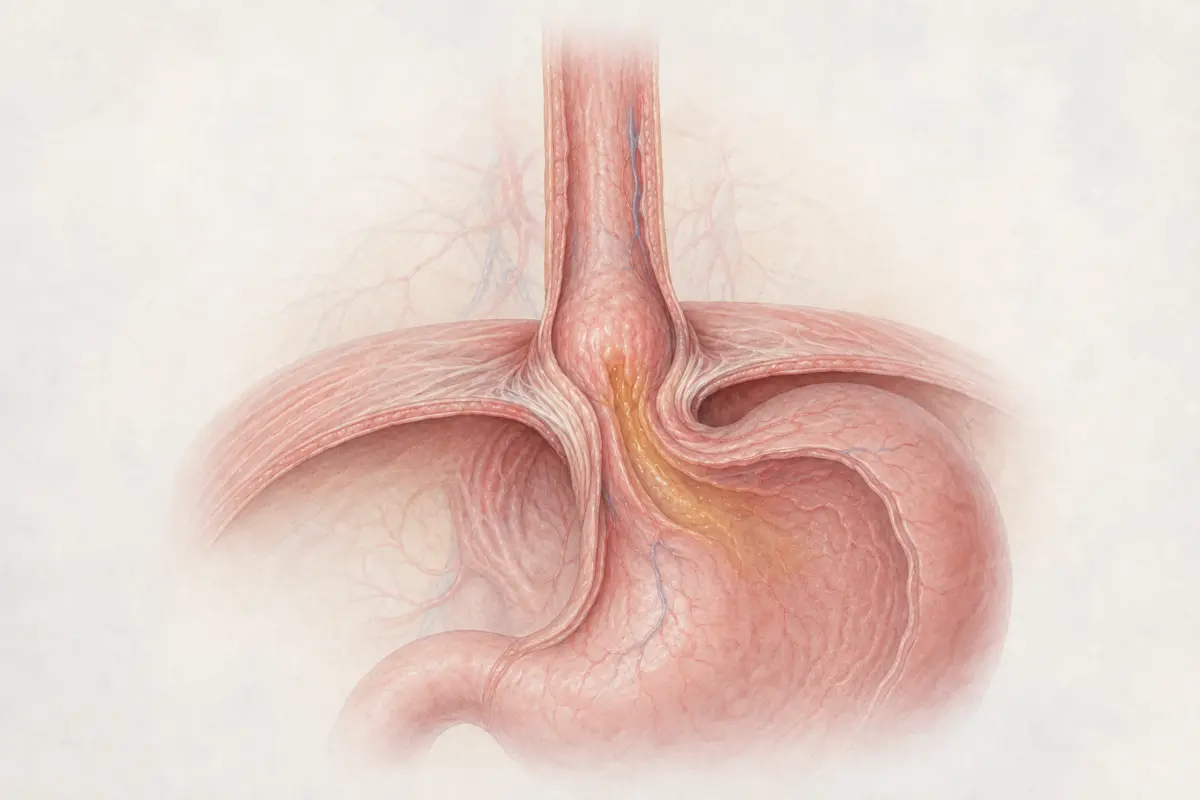
04
Reflux- & Oberbauchchirurgie
Minimalinvasive und robotische Eingriffe bei Reflux, Zwerchfellhernien und Erkrankungen des Oberbauchs.
05
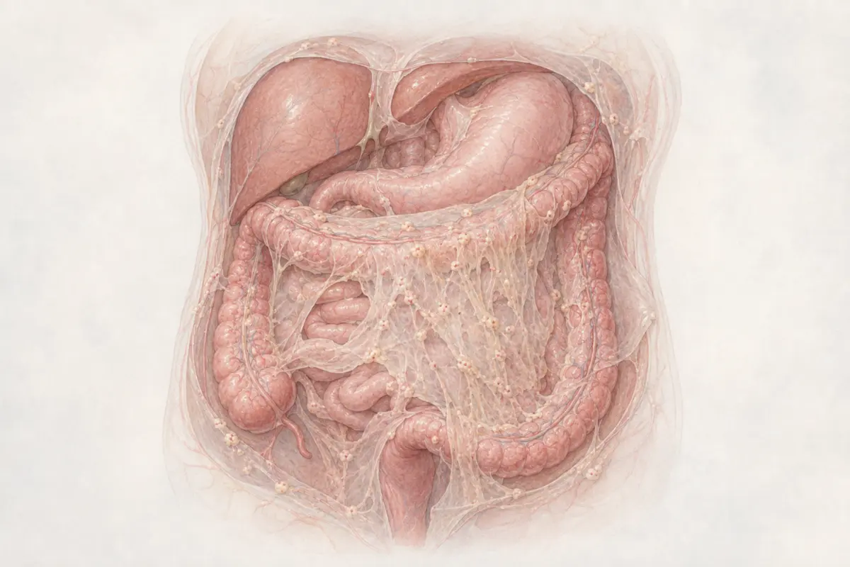
Peritonealkarzinose
Spezialisierte operative Behandlung mit zytoreduktiver Chirurgie sowie HIPEC, PIPAC und PITAC.
06
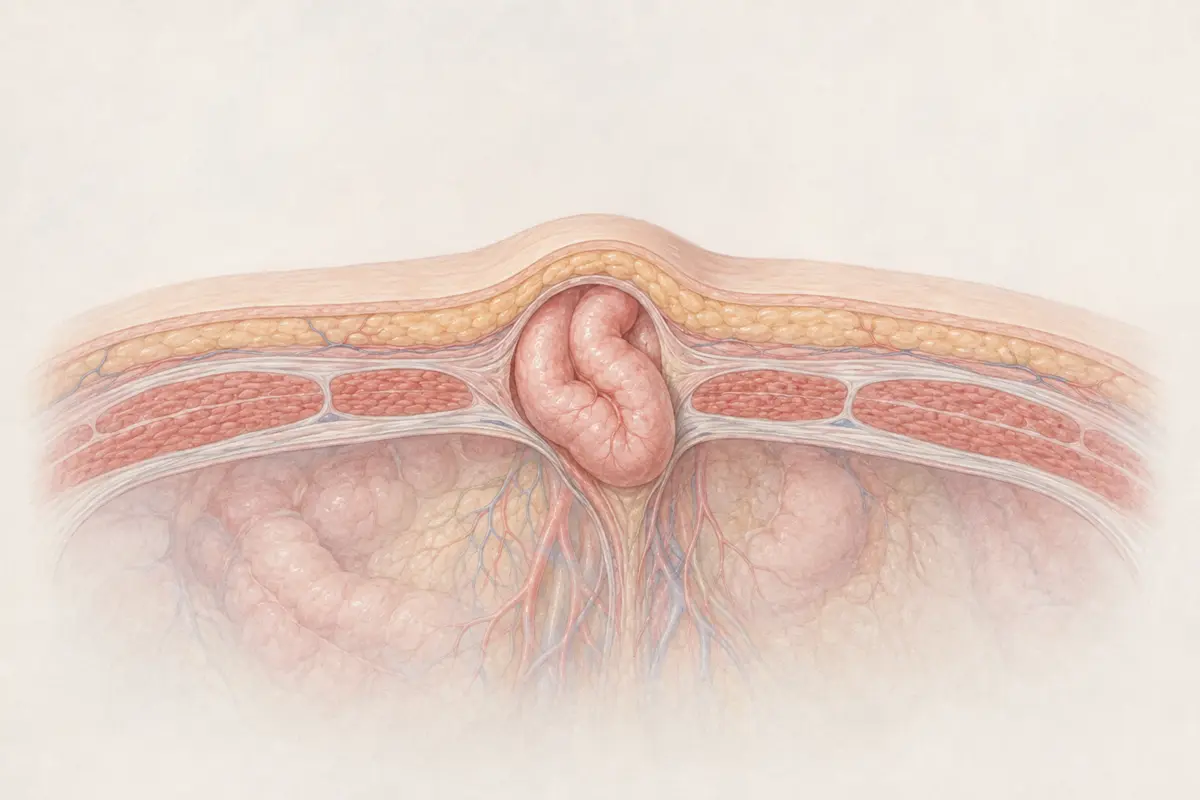
Hernien- & Bauchdeckenchirurgie
Robotische und minimalinvasive Versorgung komplexer Leisten-, Narben- und Bauchwandhernien.
07
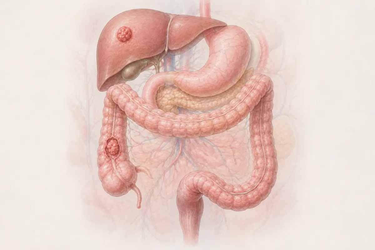
Onkologische Chirurgie
Operative Behandlung bösartiger Erkrankungen der Bauchorgane auf Grundlage interdisziplinärer Tumorkonferenzen und individuell abgestimmter Therapiekonzepte.
08
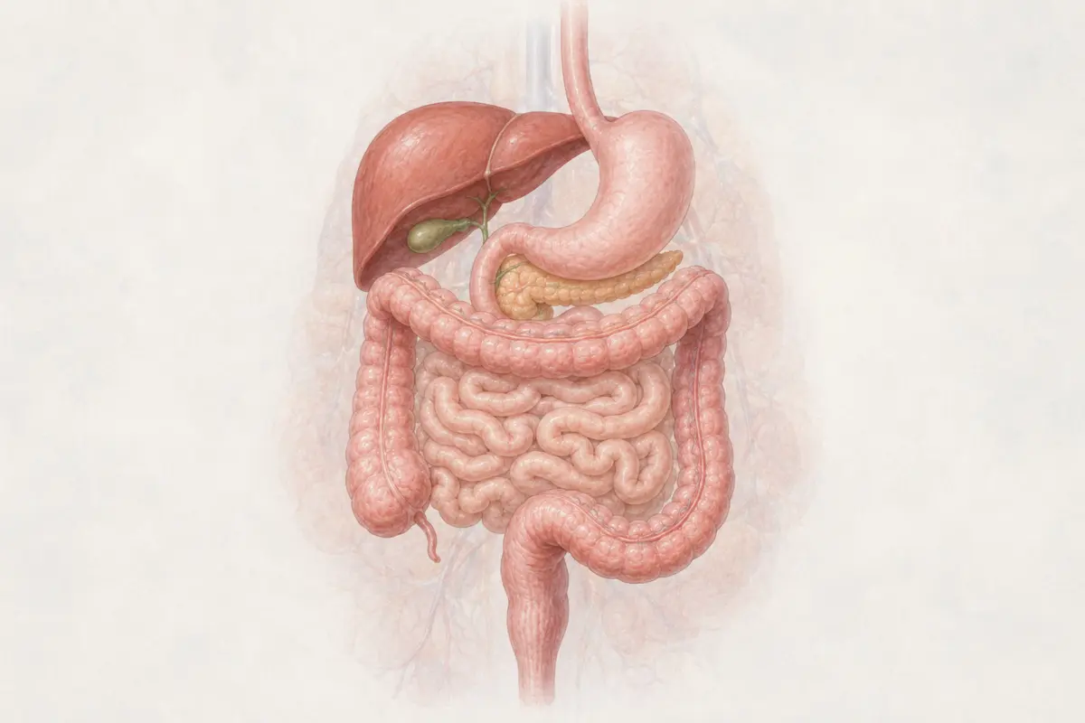
Gastrointestinale Chirurgie
Offene, minimalinvasive und robotische Behandlung gut- und bösartiger Erkrankungen von Speiseröhre, Magen, Dünndarm, Kolon und Rektum.
ROBOTIC & MINIMALLY INVASIVE SURGERY
Robotic Colorectal Surgery
Precise treatment of benign, inflammatory and oncological diseases of the colon, rectum and pelvic floor—individually planned and coordinated across disciplines.
BENIGN & INFLAMMATORY
Diseases of the colon and rectum
Each condition requires its own diagnostic and treatment strategy. Surgery is not always necessary, but may become important when complications or persistent symptoms occur.
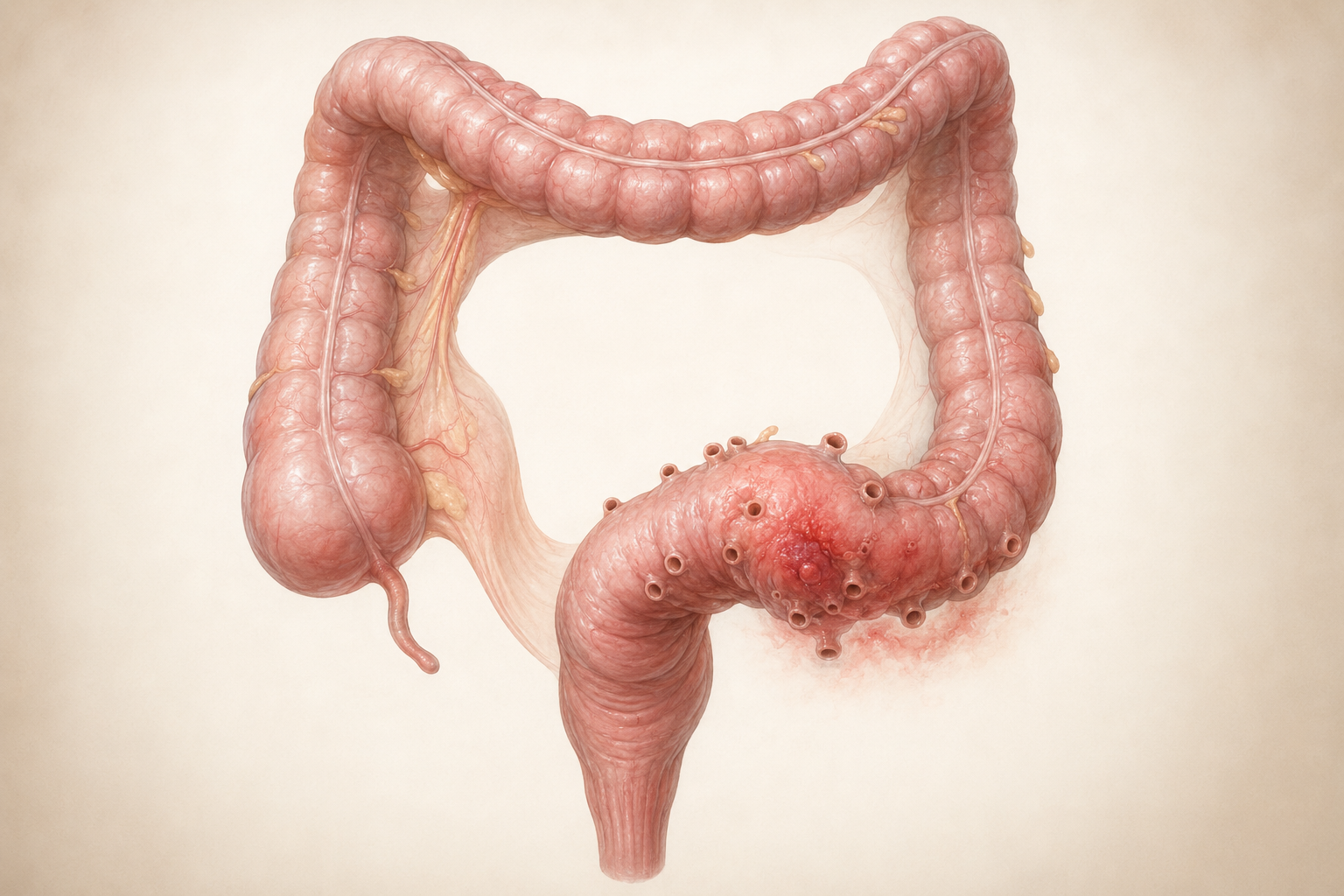
01
Diverticular Disease & Diverticulitis
What is it?
Diverticula are small pouches in the wall of the colon, most commonly in the sigmoid colon. When they become inflamed, this is called diverticulitis. Complications may include abscesses, fistulas, narrowing or perforation.
Treatment
Uncomplicated cases can often be managed without surgery. Image-guided drainage may be required for an abscess. Surgery is considered particularly for perforation, peritonitis, fistulas, significant narrowing or recurrent complicated disease.
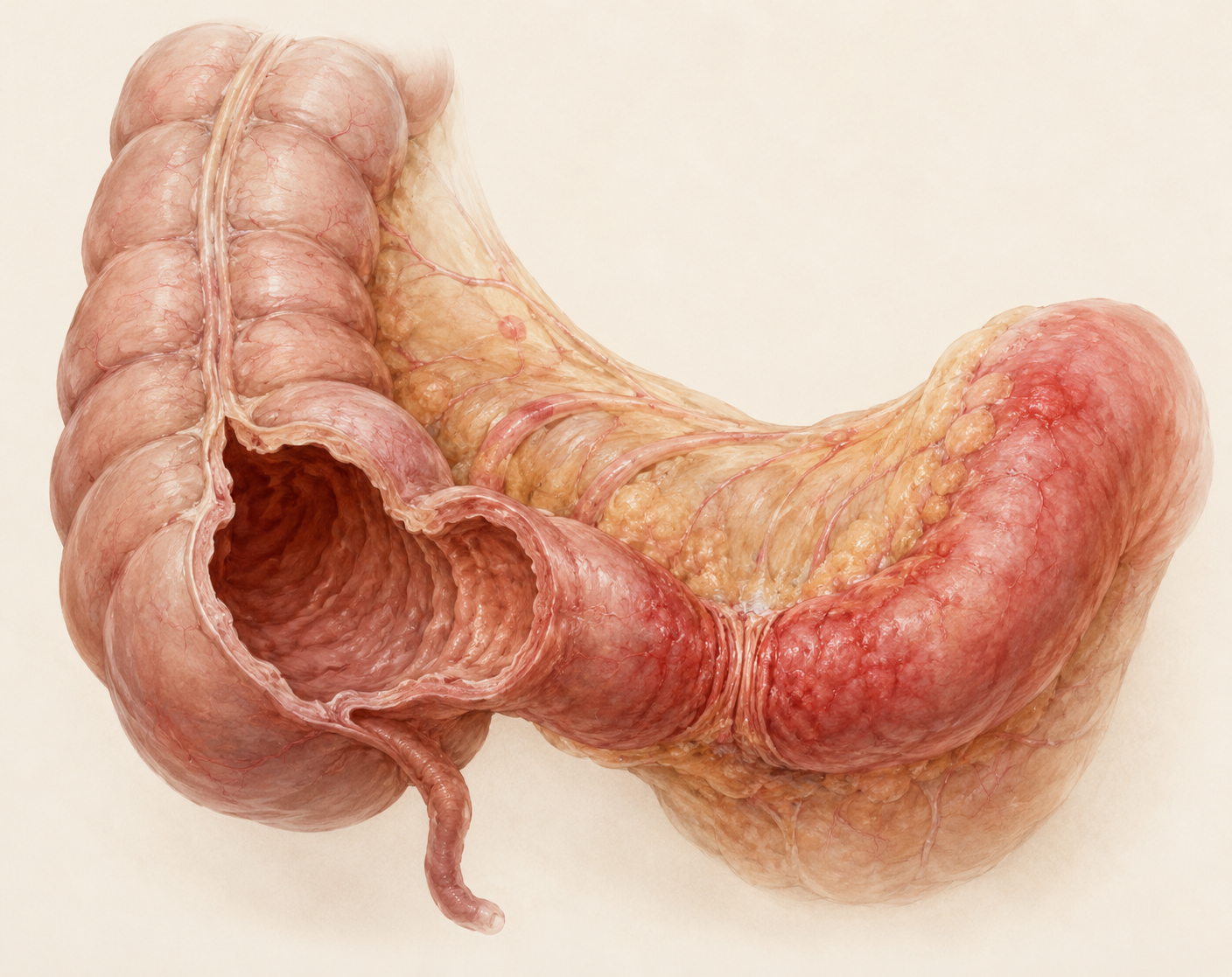
02
Crohn's Disease
What is it?
Crohn's disease is a chronic inflammatory bowel disease that can affect different parts of the digestive tract. Typical complications include strictures, fistulas, abscesses and bowel obstruction.
Treatment
Medical control of inflammation is central. Surgery may be needed for strictures, fistulas, abscesses, perforation or inadequate response to medication. It treats the complication but does not permanently cure the underlying disease.
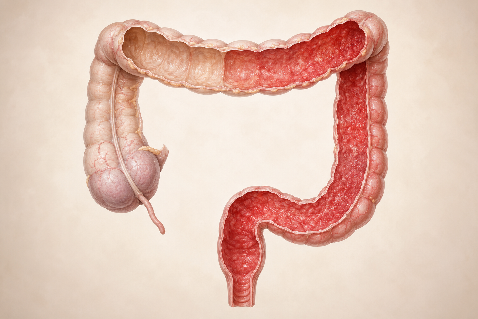
03
Ulcerative Colitis
What is it?
Ulcerative colitis is a chronic inflammatory disease of the colon. Inflammation typically begins in the rectum and may extend continuously through further sections of the colon.
Treatment
Depending on disease activity, anti-inflammatory, immunomodulating or targeted medicines are used. Surgery may be necessary for treatment-resistant disease, severe complications, precancerous changes or cancer. An ileoanal pouch reconstruction may be possible for suitable patients.
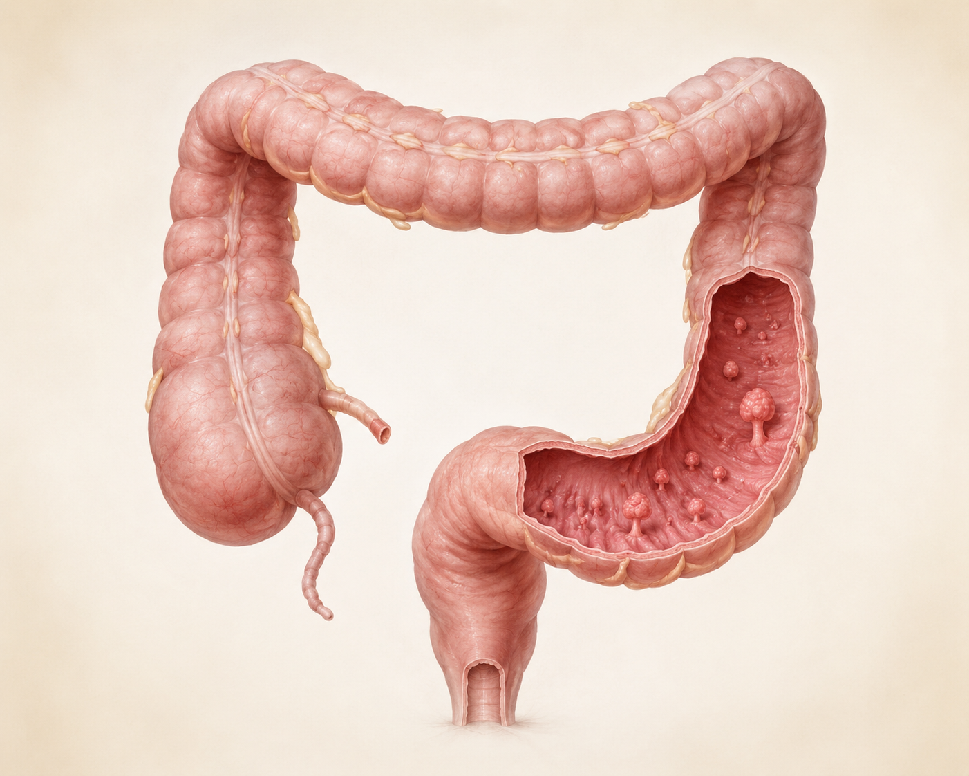
04
Colorectal Polyps & Polyposis
What is it?
Polyps are changes in the lining of the colon or rectum. Many remain benign, but certain adenomas or serrated lesions can develop into cancer over time. In inherited polyposis syndromes, numerous polyps may arise at a young age.
Treatment
Most polyps are removed during colonoscopy and examined microscopically. Surgery may be required when complete endoscopic removal is not possible, cancer is suspected or an inherited syndrome carries a high cancer risk.
05
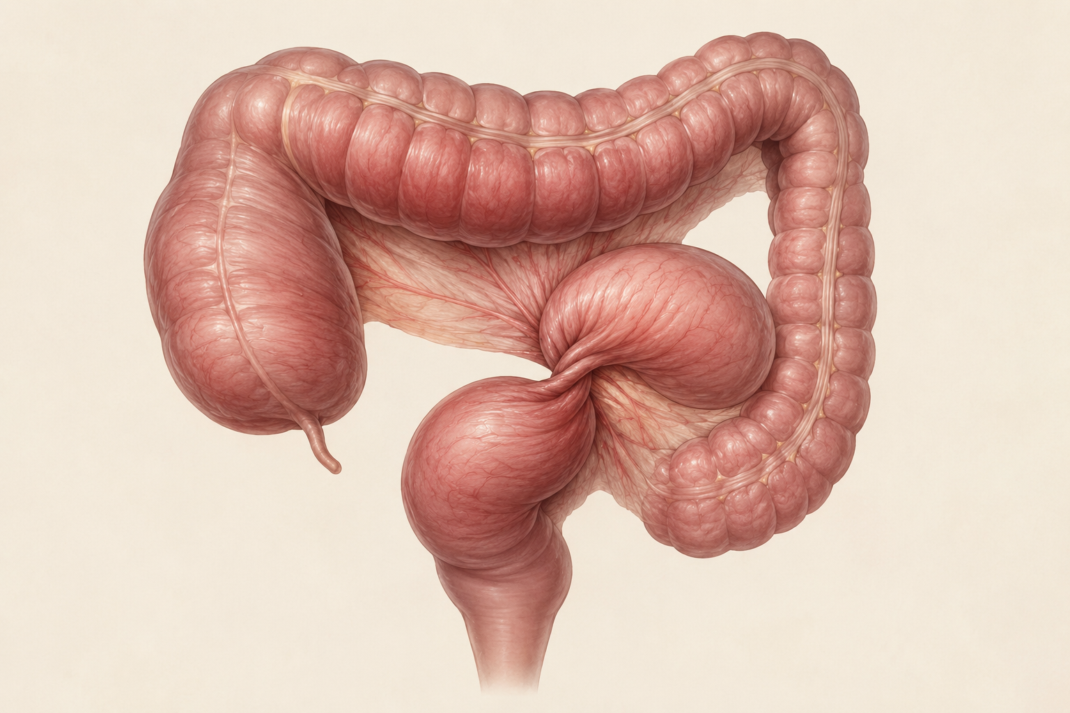
Colon Volvulus & Benign Obstruction
What is it?
In a colon volvulus, a segment of the colon twists around itself, potentially blocking the bowel and its blood supply. Benign obstruction may also result from strictures, adhesions or inflammatory changes.
Treatment
Complete bowel obstruction is a medical emergency. A sigmoid volvulus can often first be decompressed endoscopically if the bowel is viable. Because recurrence is common, planned surgery often follows. Urgent surgery is required if blood supply is compromised, perforation or peritonitis occurs.
06
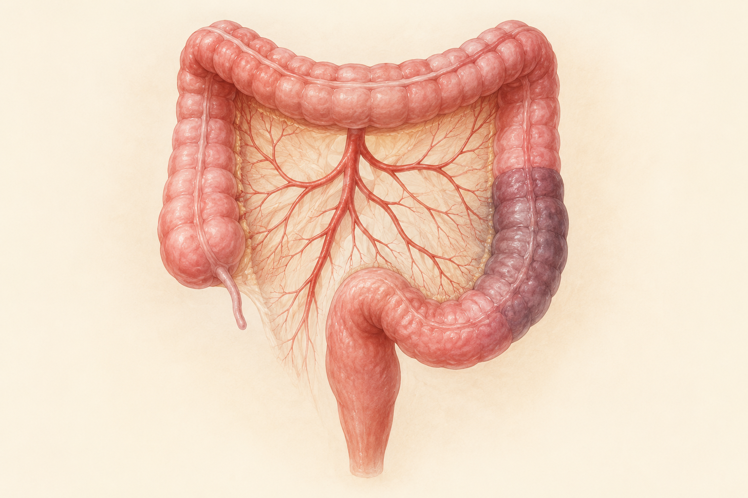
Ischemic Colitis
What is it?
In ischemic colitis, part of the colon temporarily or permanently receives insufficient blood flow. The condition may begin suddenly with abdominal pain and bloody stool.
Treatment
Milder cases may be managed with fluids, bowel rest, treatment of the cause and close monitoring. Bowel necrosis, perforation, peritonitis or ongoing deterioration require urgent surgery.
07
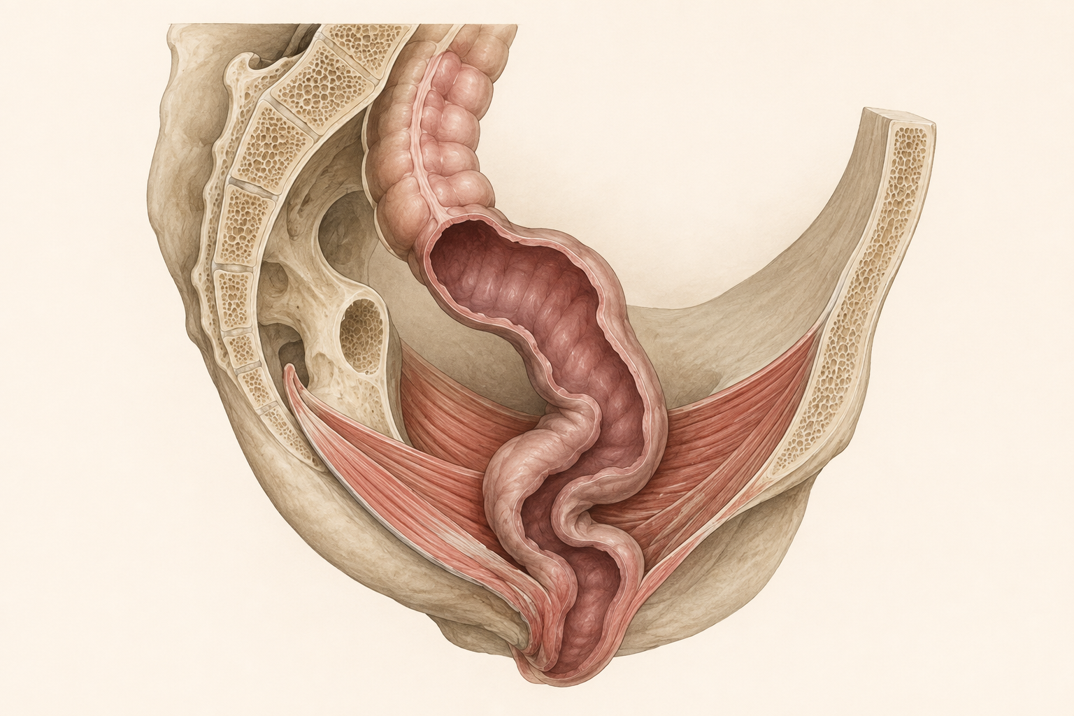
Rectal Prolapse
What is it?
In rectal prolapse, the wall of the rectum protrudes partially or completely. Associated symptoms may include difficulty emptying, incontinence, mucus discharge or bleeding.
Treatment
Bowel regulation and pelvic-floor therapy can relieve symptoms, but complete prolapse is usually treated surgically. Depending on overall health and anatomy, abdominal, minimally invasive or perineal procedures may be considered.
ONCOLOGICAL DISEASES
Colon and Rectal Cancer
Both tumour types are colorectal cancers, but they differ especially in local staging, treatment before surgery and operative strategy.
08 · COLON
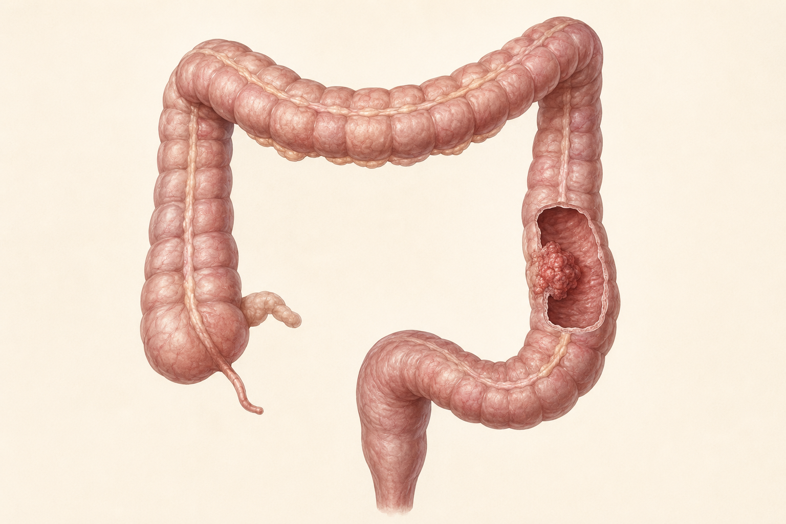
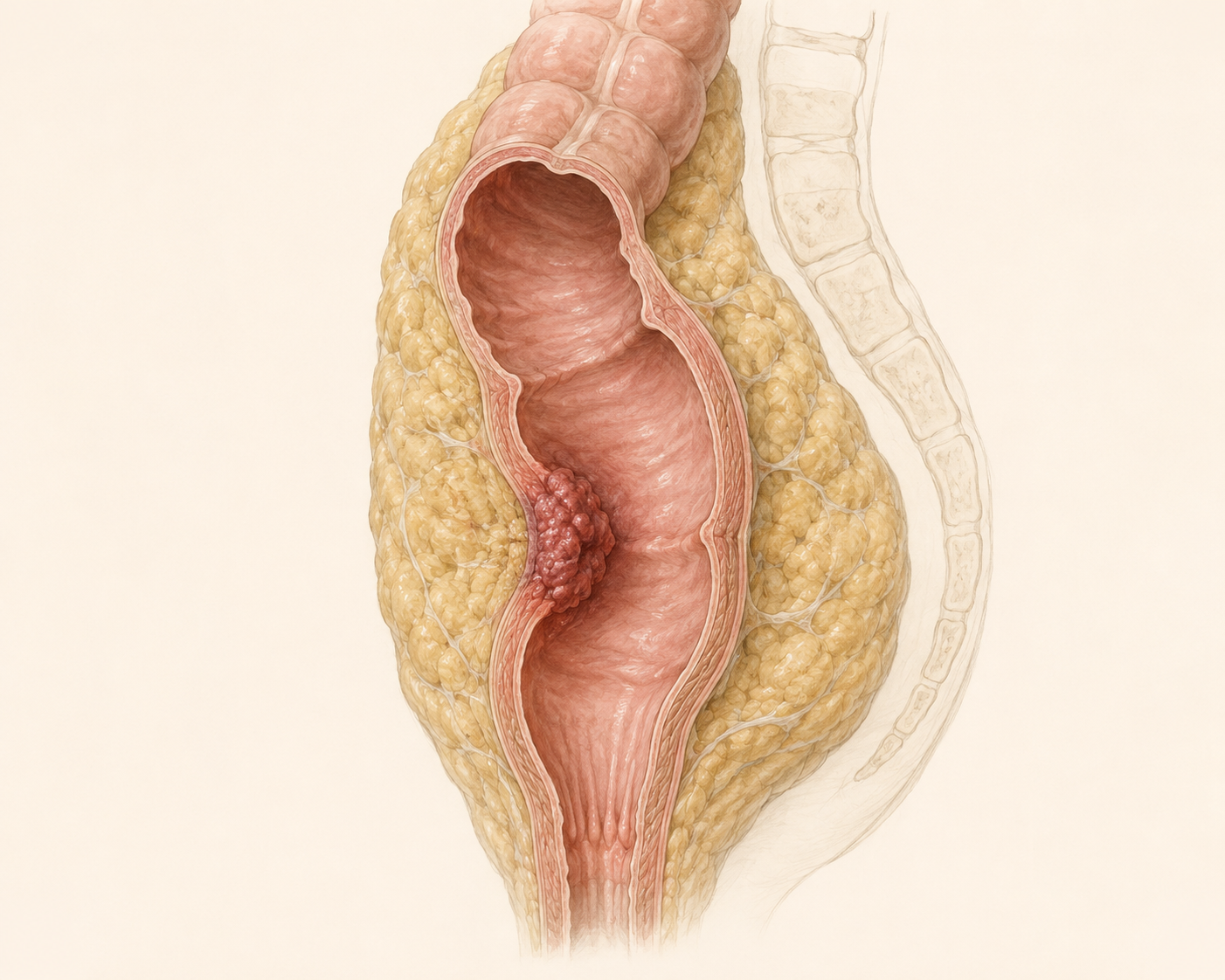
Colon Cancer
What is it?
Colon cancer is a malignant disease of the large bowel that often develops from precursors such as adenomas. Early tumours may cause no symptoms. Possible signs include blood in the stool, altered bowel habits, anaemia, weight loss or obstruction.
Treatment
For localised tumours, oncological removal of the affected bowel segment and its regional lymph nodes is the central treatment. Additional chemotherapy depends on stage, risk factors and molecular features.
09 · RECTUM
Rectal Cancer
What is it?
Rectal cancer develops in the final part of the large bowel. Because of the confined anatomy of the pelvis, its treatment differs substantially from that of colon cancer.
Treatment
Favourable early tumours may be removed locally in carefully selected patients. For locally advanced disease, chemotherapy and radiation are often combined before surgery. Surgery follows oncological principles, usually including complete removal of the mesorectum.
US GUIDELINES & CANCER STATISTICS
Diagnosis, Numbers and Treatment Pathways
Population-based estimates; not an individual prognosis. American Cancer Society ↗
Localised91.3%
Regional75.2%
Distant16.9%
Relative five-year survival rates across large patient groups. These figures are not an individual prognosis.
NCI SEER ↗Colon and rectum
- Medical history and physical examination
- Complete colonoscopy with biopsy
- CEA and further laboratory tests
- CT of chest, abdomen and pelvis
- MMR/MSI testing and molecular diagnostics when indicated
- Assessment for hereditary cancer syndromes
- Multidisciplinary tumour board
Precise local staging
- Digital rectal examination
- Rigid proctoscopy to determine tumour height
- High-resolution pelvic MRI
- Assessment of mesorectum and circumferential margin
- Relationship to the sphincter complex
- Endorectal ultrasound in selected early tumours
Stage-based treatment
EARLY
Some very early cancers within polyps can be removed completely endoscopically. Histological risk features and secure complete removal are decisive.
I
Oncological removal of the affected bowel segment and regional lymph nodes.
II
Surgery is the foundation; additional chemotherapy is considered according to individual risk factors and molecular features.
III
Adjuvant chemotherapy is generally recommended after oncological surgery.
IV
Individual planning with systemic therapy, targeted medicines or immunotherapy and—in selected patients—surgery or local treatment of metastases.
Multimodal treatment
EARLY
Small favourable tumours may be removed locally in carefully selected patients. Higher-risk disease requires oncological rectal resection.
TNT
Total neoadjuvant therapy is frequently used for locally advanced disease: chemotherapy and radiation are delivered before surgery in an individually selected sequence.
TME
Surgery aims for complete tumour removal, total mesorectal excision, clear margins and—when oncologically possible—preservation of the sphincter.
W&W
After a complete clinical response, a closely monitored watch-and-wait strategy may be discussed for selected patients in specialised centres.
Medical basis
This structure follows current US professional and patient information. Individual treatment is determined after complete diagnostic evaluation and multidisciplinary tumour-board review.
ROBOTIC COLORECTAL SURGERY
Precision in complex procedures
Robotic surgery can be particularly useful for procedures in the confined pelvis or for complex reconstructions. The system provides high-definition three-dimensional vision and precise instrument movement.
The robot does not operate independently. Every movement is fully controlled by the surgeon. The operative approach is selected according to the disease, anatomy, previous surgery and the individual medical situation.
01Colon and rectal cancer
02Diverticular disease
03Inflammatory bowel disease
04Rectal prolapse & pelvic floor
05Complex reconstructions & repeat surgery
PERSONAL & MULTIDISCIPLINARY PLANNING
Every treatment begins with a precise diagnosis.
For cancer, treatment is planned with gastroenterology, oncology, radiology, radiation oncology and pathology in a multidisciplinary tumour board.
The information on this page is intended for general patient education and does not replace a personal medical examination or individual treatment recommendation.
Services & Specialties
SPECIALIZED ONCOLOGICAL SURGERY
Peritoneal Surface Malignancies
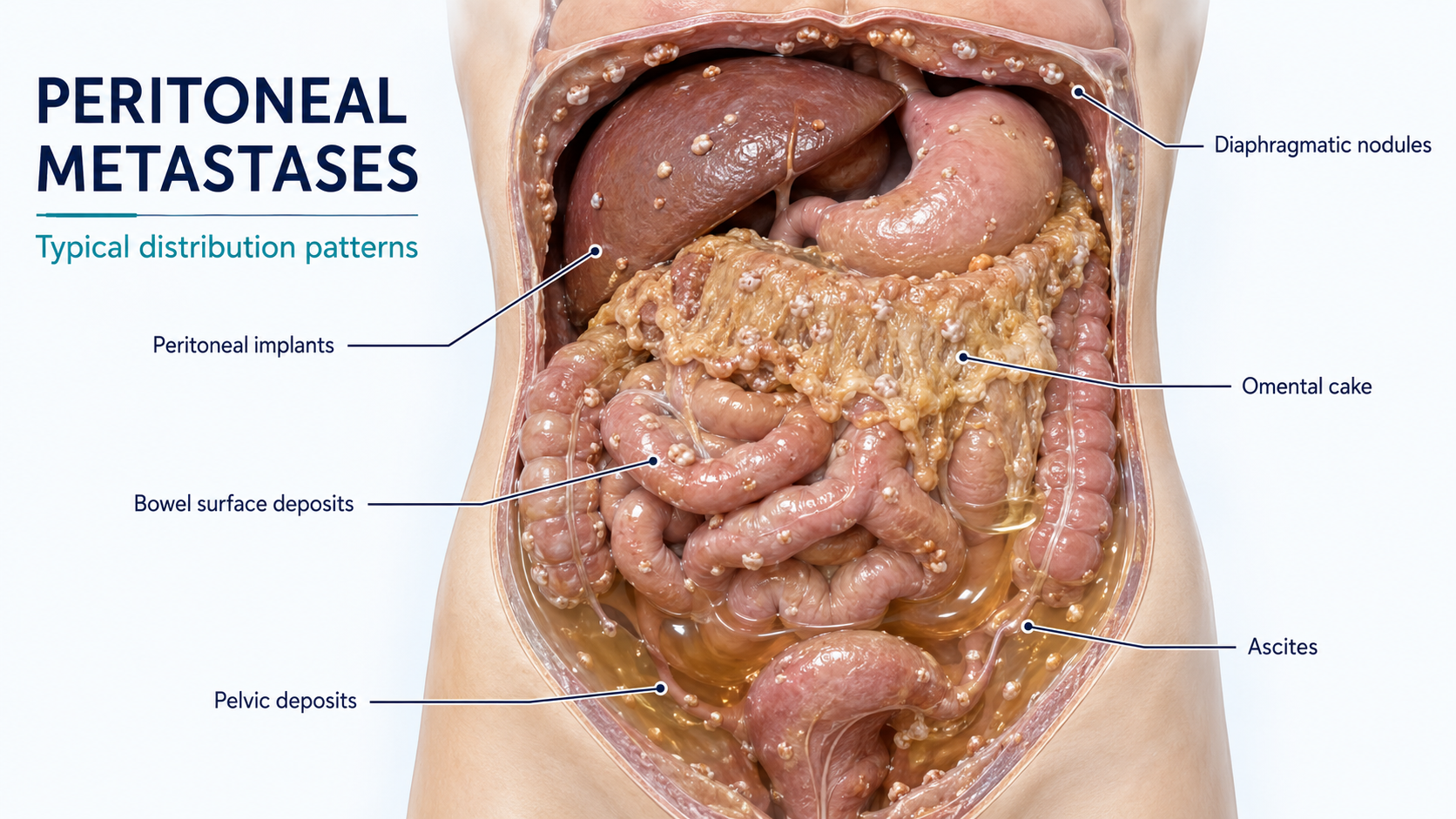
Peritoneal carcinosis occurs when cancer spreads to the membrane lining the abdominal cavity. Modern treatment may combine carefully selected surgery with regional chemotherapy delivered directly to the compartment where the disease is located.
UNDERSTANDING PERITONEAL CARCINOSIS
When cancer spreads within the abdominal cavity
Cancer cells may settle on the peritoneal lining and involve several abdominal organs. Because the peritoneum forms a distinct anatomical compartment, treatment often requires a strategy beyond systemic chemotherapy alone.
Where can it originate?
Peritoneal disease can arise from several primary cancers. The biology of the original tumour remains central to selecting the safest and most appropriate treatment.
Colorectal cancerGastric cancerAppendiceal cancerOvarian cancerMesotheliomaOther malignancies
BEFORE TREATMENT
A structured assessment guides every decision
Imaging, staging procedures and sometimes diagnostic laparoscopy help determine disease extent and whether a regional or operative approach is appropriate.
Review the diagnosis
Pathology, tumour origin, previous treatment and the current clinical situation are reviewed.
Map disease extent
Cross-sectional imaging and, where appropriate, diagnostic procedures assess distribution and resectability.
Assess fitness
General health, organ function, nutritional status and ability to recover from treatment are considered.
Multidisciplinary plan
Surgical oncology, medical oncology, radiology and pathology contribute to an individualized recommendation.
OPERATIVE & REGIONAL TREATMENT
Cytoreductive surgery and HIPEC
These treatments are distinct but may be combined in selected patients: surgery addresses visible tumour deposits, while HIPEC exposes remaining peritoneal surfaces to heated chemotherapy during the same operation.
01
CYTOREDUCTIVE SURGERY
Removing visible peritoneal disease
Cytoreductive surgery (CRS) is an extensive abdominal operation designed to remove as much visible disease as safely possible while preserving essential organ and bowel function.
- Systematic assessment of the abdomen and pelvis
- Removal of peritoneal nodules, involved peritoneum and omental disease
- Organ resection or reconstruction only when required by tumour location
- Careful balance between completeness of cytoreduction and functional preservation
02
IMPORTANT CONSIDERATIONS
Major surgery requires careful selection
CRS involves substantial tissue manipulation and a recovery period of several weeks. Potential complications include bleeding, infection and problems involving bowel reconstructions. Preoperative preparation, experienced multidisciplinary care and close postoperative monitoring are essential.
View further clinical detail about CRS
How it works
The surgeon explores all abdominal regions, identifies disease on the peritoneum and organ surfaces, and removes affected tissue where this can be achieved safely. Depending on disease location, this may include parts of the colon, small bowel, ovaries, uterus, spleen or other structures.
Treatment goal
The objective is complete or near-complete macroscopic cytoreduction while maintaining adequate bowel length and function. The realistic goal and expected benefit vary by tumour biology and disease distribution.
02
HYPERTHERMIC INTRAPERITONEAL CHEMOTHERAPY
Heated chemotherapy delivered directly to the abdomen
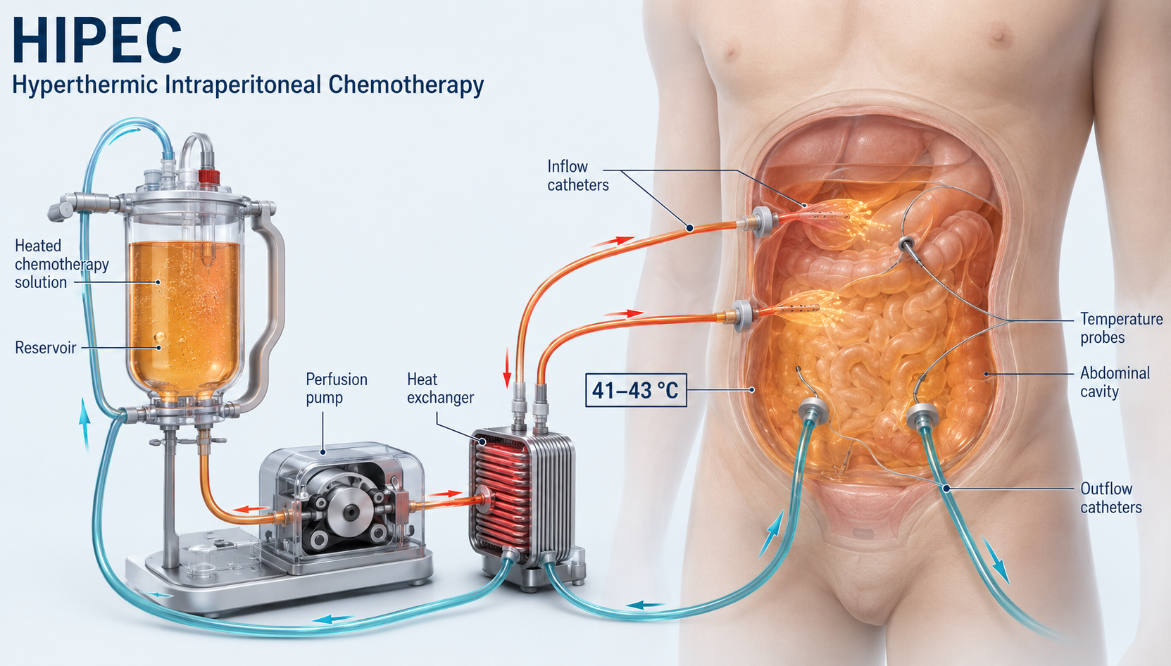
HIPEC circulates a heated chemotherapy solution through the abdominal cavity, usually immediately after cytoreductive surgery. Temperature, duration and medication depend on tumour type and the centre's protocol.
- Regional delivery creates a high local drug concentration
- Heat may enhance tissue penetration and cytotoxic activity
- Continuous circulation distributes the solution over peritoneal surfaces
- HIPEC is a single intraoperative treatment, not a substitute for all systemic therapy
View further clinical detail about HIPEC
How it works
After cytoreduction, inflow and outflow catheters circulate warmed perfusate through the abdominal cavity. The team monitors temperature and distribution before the solution is removed and the operation is completed.
Why local delivery matters
The peritoneal compartment permits high local exposure while systemic absorption is comparatively limited. The expected value of HIPEC depends on the primary tumour, completeness of surgery, regimen and individual disease characteristics.
PRESSURIZED AEROSOL THERAPY
PIPAC and PITAC
Pressurized aerosol techniques deliver regional chemotherapy through minimally invasive access. They do not remove tumour tissue and should be considered within a specialized oncological treatment plan.
03
PRESSURIZED INTRAPERITONEAL AEROSOL CHEMOTHERAPY
PIPAC: regional treatment through laparoscopy
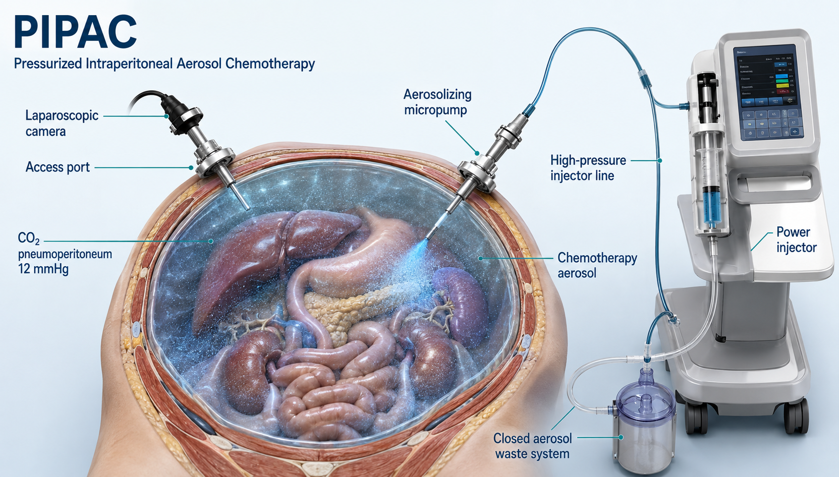
PIPAC uses a specialized nebulizer to distribute chemotherapy as a fine aerosol in the pressurized abdominal cavity. It is performed laparoscopically in an operating-room setting, generally under general anaesthesia.
- Small laparoscopic access points and camera visualization
- Controlled aerosol delivery within a closed system
- Repeatable treatment in selected protocols
- Potential use for unresectable disease, palliation or within downstaging strategies
View further clinical detail about PIPAC
Aerosol and pressure
The aerosol format and controlled pressure are intended to improve distribution over exposed peritoneal surfaces. Treatment remains confined to the abdominal cavity and is carried out with occupational safety measures and a closed waste system.
Role in treatment
PIPAC may be discussed when major cytoreductive surgery is not appropriate, as part of symptom-oriented treatment, or in selected multimodal protocols. Availability, medication and treatment intervals vary between specialist centres.
PIPAC
Targets disease on the peritoneal surfaces through laparoscopic access to the abdomen.
01Laparoscopic visualization and staging
02Controlled aerosol chemotherapy delivery
03Potentially repeatable in selected treatment plans
PITAC
Extends the aerosol concept to the pleural cavity for selected patients with malignant pleural disease. Access is obtained by thoracoscopy, followed by controlled aerosol delivery and drainage.
01Thoracoscopic access to the pleural space
02Pressurized regional chemotherapy exposure
03Protocol-driven treatment at selected specialist centres
PIPAC versus PITAC
The same regional aerosol principle, applied to different anatomical compartments.
Aspect
PIPAC
PITAC
Target
Peritoneal cavity (abdomen)
Pleural cavity (thorax)
Access
Laparoscopy
Thoracoscopy
Experience
Growing clinical and research experience
More limited; evolving evidence
Current role
Specialized, protocol-dependent option
Primarily investigational / protocol-driven
CHOOSING THE RIGHT APPROACH
No single treatment fits every patient
Treatment selection integrates tumour biology, disease extent and location, resectability, overall health, previous therapies and each patient's goals.
CRS with or without HIPEC
May be considered when peritoneal disease appears technically removable and the patient is fit for a major multimodal procedure.
- Potentially resectable disease
- Adequate health and organ function
- Careful specialist assessment
- Commitment to recovery and follow-up
PIPAC
May be discussed when major cytoreduction is not appropriate or as part of a specialized multimodal treatment strategy.
- Regional therapy without tumour removal
- Protocol-dependent repeat treatments
- Specialized equipment and expertise
- Evidence and availability continue to evolve
Medical oncology
Systemic chemotherapy, targeted treatment or immunotherapy remains central for many patients and may be combined with or used instead of regional procedures.
- Tumour-specific treatment plan
- Sequencing before or after surgery
- Response assessment over time
- Joint surgical-oncology decision
OUR APPROACH
Clear information. Honest expectations. Individual planning.
Peritoneal carcinosis is complex. Our aim is to explain the available options, coordinate specialist assessment and define a realistic treatment strategy aligned with the patient's disease, health and personal priorities.
01
Thorough assessmentReview of imaging, pathology, prior treatment and disease distribution.
02
Multidisciplinary collaborationClose coordination with medical oncology, imaging and pathology specialists.
03
Balanced consultationTransparent discussion of expected benefit, risks, alternatives and recovery.
04
Individual treatment planIntegration of operative, regional and systemic treatment where appropriate.
This information supports, but does not replace, individual medical advice. Treatment suitability can only be determined after personal consultation and review of the complete medical record.


PD. Dr. Omar Thaher
Consultant General, Visceral & Robotic Surgeon
German Board-Certified · 15 Years of Surgical Experience
DA VINCI
ÜBER MICH
Moderne Chirurgie mit Präzision, Erfahrung und Menschlichkeit.
Als Facharzt für Viszeralchirurgie und spezielle Viszeralchirurgie liegt mein besonderer Schwerpunkt auf der roboterassistierten und minimalinvasiven Behandlung komplexer Erkrankungen des Bauchraums.
Meine operative Tätigkeit umfasst insbesondere die kolorektale und Beckenbodenchirurgie, Reflux- und Oberbauchchirurgie, komplexe Hernienchirurgie sowie die Behandlung der Peritonealkarzinose. Ergänzt wird die klinische Arbeit durch universitäre Lehre, wissenschaftliche Forschung und den Aufbau moderner robotischer Operationsprogramme.
„Bei aller Spezialisierung und dem Einsatz modernster Techniken steht der Mensch als Individuum im Mittelpunkt meines Denkens und Handelns.“
Facharztqualifikationen
Viszeralchirurgie, spezielle Viszeralchirurgie und Proktologie
Roboterchirurgische Expertise
Fortgeschrittene Erfahrung mit da Vinci
Wissenschaft & Lehre
Habilitation, universitäre Lehre und Publikationen
Aktuelle Aufgabe
Aufbau der Roboterchirurgie mit Schwerpunkt kolorektale Chirurgie
ABOUT ME
PD. Dr. Omar Thaher
Consultant General, Visceral & Robotic Surgeon
German Board-Certified · Special Visceral Surgery & Proctology · 15 Years of Surgical Experience
German board certification and 15 years of operative experience in general, visceral and specialised visceral surgery form the foundation of my clinical work.
The principal clinical focus lies in robotic and minimally invasive surgery, complex abdominal procedures and the surgical treatment of benign and malignant diseases of the gastrointestinal tract. Operative expertise is combined with clinical leadership, academic research and surgical education.
“Modern surgery requires precision, experience and innovation. Yet even with the most advanced technology, the individual patient must always remain at the centre of every medical decision.”
MY APPROACH TO PATIENT CARE
Individual planning. Clear communication. Modern surgical care.
Every patient and every clinical situation is different. Careful evaluation, clear communication and an individually tailored treatment plan therefore form the foundation of my work.
Evidence-based careCurrent medical evidence and modern, patient-centred techniques provide the basis for every treatment decision.
Individual choice of procedureMinimally invasive and robotic procedures are used whenever they are medically appropriate for the individual clinical situation.
Patient at the centreTechnical precision is valuable when it supports safety, structured recovery and long-term well-being.
CLINICAL EXPERTISE
Specialised surgical expertise
Extensive operative experience, modern techniques and multidisciplinary decision-making shape the treatment of complex abdominal conditions.
Robotic & Minimally Invasive Surgery
Extensive experience in robotic and minimally invasive procedures using the da Vinci surgical system, including complex operations of the upper gastrointestinal tract, colon and rectum, abdominal wall and pelvic floor.
Oncological Surgery
Surgical care for malignant diseases of the abdominal organs. Oncological procedures are planned within a multidisciplinary framework and adapted to the individual clinical situation.
Gastrointestinal Surgery
Open, minimally invasive and robotic treatment of benign and malignant diseases of the oesophagus, stomach, small intestine, colon and rectum.
Peritoneal Surface Malignancies
A particular clinical focus is the treatment of peritoneal carcinomatosis, including cytoreductive surgery and advanced procedures such as HIPEC, PIPAC and PITAC.
Colorectal Surgery
Treatment of benign and malignant diseases of the colon and rectum using established open, minimally invasive and robotic techniques.
Hernia & Abdominal Wall Surgery
Treatment of primary, recurrent, incisional and complex abdominal wall hernias using individually selected open, minimally invasive or robotic techniques.
Proctological Surgery
Modern diagnostic and surgical treatment of common and complex proctological conditions, with particular attention to preserving function and supporting recovery.
PROFESSIONAL CAREER
Clinical experience and surgical leadership
The professional path combines comprehensive surgical training with increasing clinical responsibility and senior leadership roles in Germany and Switzerland.
- 01
Lead Physician | Department of Surgery
Spital HerisauHerisau · Switzerland
- 02
Lead Senior Consultant & Deputy Head of Department
Department of General and Visceral SurgeryUniversity Hospital Herne · Ruhr University Bochum
Herne · Germany - 03
Managing Senior Consultant & Deputy Head of Department
Department of General and Visceral SurgeryUniversity Hospital Herne · Ruhr University Bochum
Herne · Germany - 04
Senior Consultant
Department of General and Visceral SurgeryUniversity Hospital Herne · Ruhr University Bochum
Herne · Germany - 05
Surgical Resident | General, Visceral & Thoracic Surgery
Klinikum Darmstadt GmbHDarmstadt · Germany
- 06
Surgical Resident | General & Visceral Surgery
Bürgerhospital Frankfurt am MainFrankfurt am Main · Germany
- 07
Surgical Resident | General, Visceral & Minimally Invasive Surgery
Klinikum Bad HersfeldBad Hersfeld · Germany
MEDICAL EDUCATION & LICENSURE
Medical training and professional recognition
Medical studies, professional licensure and recognition of the medical diploma in Switzerland form the foundation of clinical practice.
Recognition of Medical Diploma in Switzerland
Medical Professions Commission – MEBEKO · Switzerland
Medical Licence to Practise – Approbation
State Medical Association of Hesse · Germany
Medical Studies – Human Medicine
Justus Liebig University Giessen · Giessen · Germany
SURGICAL LEADERSHIP & QUALITY
Reliable structures for better surgical care
Modern surgical care requires more than technical expertise in the operating room. Reliable clinical structures, interdisciplinary collaboration and clearly defined treatment pathways are essential for patient safety and consistent treatment quality.
- Development of multidisciplinary surgical teams
- Establishment of clinical and organisational standards
- Support of certification processes for specialised surgical centres
- Introduction of modern operative procedures and treatment concepts
- Quality management and structured evaluation of clinical outcomes
- Training and supervision of surgical colleagues
TEACHING, EXAMINATION & TRAINING AUTHORISATION
Sharing knowledge. Building responsibility.
Academic teaching combines theoretical knowledge with clinical decision-making, practical surgical skills, risk management and perioperative safety. Structured teaching and individual supervision support the gradual assumption of professional responsibility.
PROFESSIONAL & SCIENTIFIC MEMBERSHIPS
DGAVGerman Society for General and Visceral Surgery
DAGGerman Obesity Society
CAADIPSurgical Working Group for Obesity and Metabolic Surgery
ACOAssociation of Surgical Oncology
EHSEuropean Hernia Society
ISSPPInternational Society for the Study of Pleura and Peritoneum
IOSInternational Obesity Society
GBDGlobal Burden of Disease
Professional exchange across disciplines and borders
DOCTORATE & HABILITATION
DOCTORATE · DR. MED.
HABILITATION
Academic qualifications
Propensity Score Matching: Sleeve Gastrectomy vs. Gastric Bypass in Older Patients Aged 60 Years and Above
Otto von Guericke University Magdeburg · Germany
Supervisor: Prof. Dr. med. Christine Stroh
The Significance and Impact of Obesity and Metabolic Surgery in the Federal Republic of Germany
Faculty of Medicine · Ruhr University Bochum · Germany
Supervisor: Prof. Dr. med. Dirk Bausch
RESEARCH & ACADEMIC WORK
Connecting clinical practice with scientific evidence
The continuous evaluation of surgical techniques, treatment pathways and clinical outcomes provides an important foundation for improving patient safety and long-term treatment quality.
- Robotic and minimally invasive surgery
- Oncological and visceral surgery
- Peritoneal surface malignancies
- Obesity and metabolic surgery
- Surgical quality and outcome research
- Digitalisation and artificial intelligence in surgery
INNOVATION & THE FUTURE OF SURGERY
Integrating technology responsibly into care
I have a strong interest in the responsible integration of modern technologies into surgical care. My work focuses particularly on robotic surgery, digital clinical processes and the potential application of artificial intelligence in preoperative preparation and postoperative patient care. My objective is to provide more precise, structured and patient-centred treatment while respecting medical ethics, data protection and patient trust.
“My goal is to combine surgical experience, scientific evidence and modern technology with personal, respectful and transparent patient care. Every treatment decision should be understandable, individually appropriate and focused on the best possible outcome for the patient.”PD. Dr. Omar Thaher
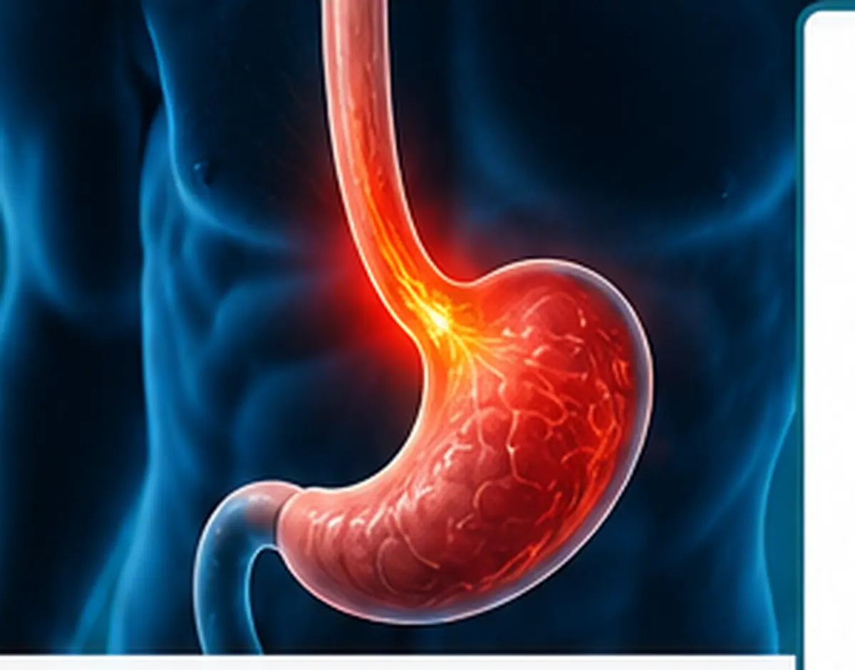
REFLUX & UPPER ABDOMINAL SURGERY
Reflux Disease & Hiatal Hernias
Modern diagnostics and individually tailored treatment for gastro-oesophageal reflux disease and hiatal hernias—from conservative care to minimally invasive reconstruction.
Reflux is more than excess stomach acid.
Gastro-oesophageal reflux disease occurs when stomach contents repeatedly flow back into the oesophagus, causing symptoms or mucosal injury. The central problem is often not acid quantity alone, but an impaired barrier at the junction between the oesophagus and stomach.
This barrier includes the lower oesophageal sphincter, the diaphragm, the angle between oesophagus and stomach, and coordinated oesophageal and gastric motility. A hiatal hernia can impair this interaction, but does not automatically mean that reflux requires treatment.
Not every hiatal hernia requires surgery. Symptoms, anatomy, objective findings and individual risk are assessed together.
Reflux oesophagitis, erosions, ulcers and peptic narrowing of the oesophagus may occur.
Long-standing reflux can lead to mucosal change that may require targeted endoscopic surveillance.
Night-time symptoms, disturbed sleep and selected reflux-associated airway symptoms can significantly affect quality of life.
RECOGNISING SYMPTOMS
Typical and less typical signs
Reflux and hiatal hernias can cause a wide range of symptoms. Careful attribution is particularly important when cough, hoarseness or swallowing difficulty are the main concern.

Heartburn
A burning sensation behind the breastbone, often after meals or while lying down.

Acid regurgitation
Sour belching or the backflow of stomach contents into the throat.

Night-time cough and hoarseness
Reflux may irritate the airways and contribute to cough or hoarseness.

Difficulty swallowing
A sensation that food is sticking in the throat or behind the breastbone.
Upper abdominal pain and fullness
Pressure, pain, bloating or early satiety in the upper abdomen.

Dental problems
Repeated acid exposure may affect tooth enamel and oral health.
Typical symptoms
- Heartburn and burning behind the breastbone
- Sour or bitter regurgitation
- Pressure or burning in the upper abdomen
- Symptoms after meals or while lying down
Other possible symptoms
- Difficulty or pain when swallowing
- Hoarseness, chronic cough or a globus sensation
- Nausea, early satiety, fullness or belching
- Mechanical pressure with larger paraoesophageal hernias
ANATOMY
Four types of hiatal hernia
The anatomical type influences symptoms, risk and treatment planning. The combination of findings and clinical circumstances is what matters.
Type I · sliding
The junction between the oesophagus and stomach slides above the diaphragm. This is the most common type.
Type II · paraoesophageal
Part of the stomach enters the chest beside the oesophagus while the junction remains in its normal position.
Type III · mixed
Sliding and paraoesophageal components occur together; larger hernias are often of this type.
Type IV · complex
In addition to the stomach, other abdominal organs may move through the enlarged hiatus into the chest.
OBJECTIVE AND STRUCTURED
Diagnostics before treatment
Symptoms alone are not sufficient for an invasive treatment decision. Endoscopy, functional testing and imaging answer different questions and are combined selectively.
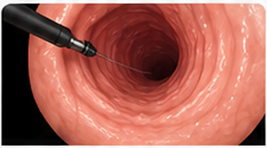
01 · ENDOSCOPY
Gastroscopy
Assesses mucosa and anatomy: reflux oesophagitis, Barrett's mucosa, narrowing, ulcers, hiatal hernia and the gastro-oesophageal junction. Biopsies are taken when indicated.
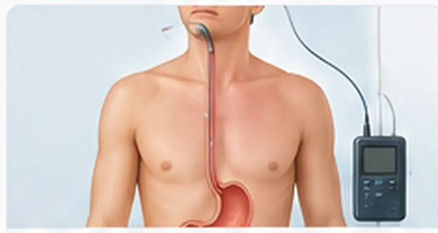
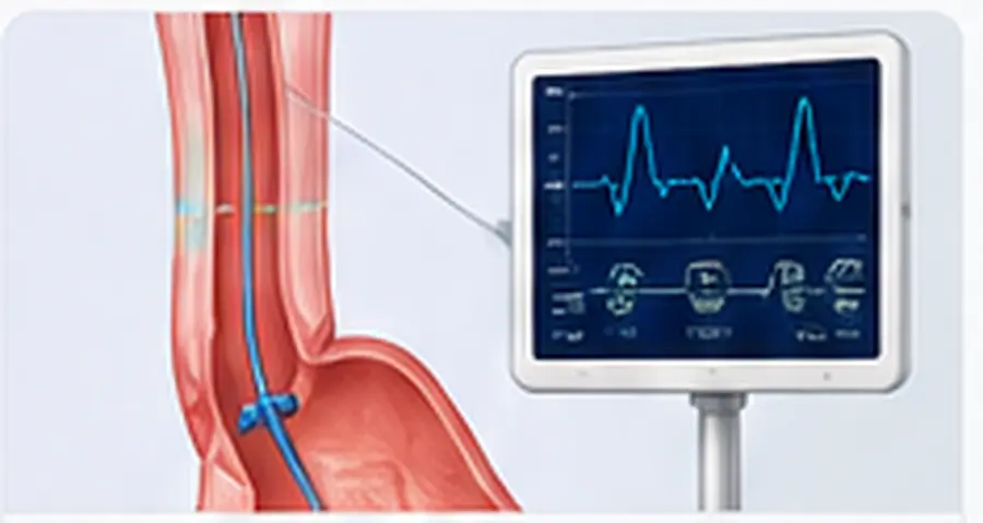
02 · REFLUX MONITORING
pH or pH-impedance monitoring
Records acid reflux for 24 hours or wirelessly over several days and—with impedance—also non-acid reflux. It is especially important when endoscopy is negative or symptoms persist.
03 · FUNCTION
High-resolution manometry
Measures pressure and movement within the oesophagus. It identifies motility disorders, helps exclude achalasia and informs selection of an appropriate operation.
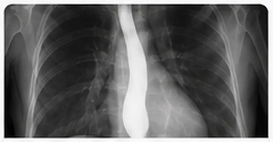
04 · ANATOMY
Contrast swallow
Shows anatomy and dynamics, hernia size and position, passage, possible shortening of the oesophagus or rotation. On its own, however, it does not prove reflux disease.
STEPWISE TREATMENT
Begin conservatively, escalate selectively
Treatment is guided by symptoms, objective evidence of reflux, anatomy and individual circumstances. The most appropriate—not simply the newest—method is what matters.
Nutrition and daily habits
Recommendations are selected individually and should avoid unnecessary restriction.
- When overweight, weight reduction can improve symptoms.
- Avoid late meals; elevate the head of the bed for night-time reflux.
- Smaller portions and personal triggers rather than rigid exclusion lists.
- Stop smoking and be cautious with very high-fat meals.
Effective acid control
Proton pump inhibitors are the main medical treatment. Timing, adherence and the lowest effective dose are reviewed regularly.
- Usually taken before a meal
- H2 blockers, alginates or antacids in selected situations
- If symptoms persist, first verify diagnosis and correct use
An inadequate response to PPIs should only be assumed after timing, dose, adherence, possible alternative diagnoses and—where required—objective reflux evidence have been reviewed.
WHEN SURGERY MAY HELP
Restore anatomy, control reflux
Surgery is considered particularly for objectively proven reflux, troublesome regurgitation, a large or symptomatic hernia and selected complications. Endoscopy, manometry and reflux testing form part of structured preoperative planning.
Hiatal reconstruction
The stomach is returned to the abdomen and the diaphragmatic crura are reconstructed behind the oesophagus. This is the central anatomical step for large hernias.
Nissen fundoplication
A complete 360° wrap reinforces the reflux barrier. It is highly effective, but may have a greater effect on swallowing, belching and gas relief.
Toupet fundoplication
A posterior partial wrap of approximately 270° may be an appropriate alternative, particularly with certain motility findings. Other partial wraps are used selectively.
INDIVIDUAL DECISION
There is no single best operation for everyone.
Choice is guided by hernia size, reflux evidence, oesophageal motility, swallowing function, previous surgery, body weight and personal priorities. Particular caution is required with unexplained dysphagia, relevant motility disorders, extraoesophageal symptoms without objective reflux evidence or high operative risk. With clinically relevant obesity, a bariatric strategy such as Roux-en-Y gastric bypass may be more appropriate.
CURRENT OPTIONS
Place newer procedures in context
Modern procedures broaden the range of options, but do not automatically replace established operations. Long-term evidence, revision options and centre experience form part of the decision.
Magnetic sphincter augmentation · LINX
For selected patients with objectively proven reflux, suitable anatomy and assessed motility. Device, erosion, removal and MRI considerations must be discussed.
TIF 2.0
Endoscopic reconstruction for selected patients with confirmed reflux, a small hernia and suitable anatomy. It is not suitable alone for larger hernias; long-term evidence is less extensive than for fundoplication.
cTIF
Combines surgical hernia repair with endoscopic TIF. The concept is plausible, but robust randomised long-term data remain limited.
RefluxStop
Available in selected European centres. Early and mid-term evidence is growing, but remains younger and less randomised than the evidence for established fundoplication procedures.
Roux-en-Y gastric bypass
An option for reflux with clinically relevant obesity or in complex revision situations. It is not a standard procedure for normal-weight reflux patients.
Other selective procedures
Stretta, endoscopic plication and other emerging approaches are considered only very selectively or within structured programmes because evidence and long-term comparative data are heterogeneous.
A new method should be judged by objective reflux evidence, suitable anatomy, multi-year outcomes, comparison with fundoplication or MSA, dysphagia and reoperation risk, and available revision options.
LARGE AND COMPLEX HERNIAS
Specialist planning for complex anatomy
With large paraoesophageal hernias, mechanical symptoms and the risk of entrapment or rotation often predominate. Surgery involves complete return of the stomach, management of the hernia sac, adequate oesophageal length and stable hiatal reconstruction.
Shortened oesophagus
In selected complex situations, a Collis lengthening procedure may be required so that the gastro-oesophageal junction rests in the abdomen without tension.
Risk of recurrence
Anatomical recurrence can occur even after careful reconstruction. Not every radiological recurrence causes symptoms or requires another operation.
Revision surgery
Repeat surgery after previous reflux or hernia procedures requires particularly precise diagnostics and should be planned in an experienced specialist centre.
BENEFITS, RISKS AND RECOVERY
Realistic goals, safe recovery
The goal is to improve heartburn and regurgitation, restore anatomy, protect the mucosa and improve quality of life. Complete freedom from symptoms or medication cannot be guaranteed.
Possible risks
- Bleeding, infection, thrombosis and anaesthetic risks
- Injury to adjacent structures or opening of the pleura
- Temporary or persistent swallowing difficulty
- Gas-bloat and reduced ability to belch or vomit
- Recurrent reflux, hernia recurrence or reoperation
- Procedure- or device-specific risks
Seek urgent help after surgery
- Increasing breathlessness, chest or upper abdominal pain
- Fever or a marked decline in general condition
- Inability to swallow liquids or persistent vomiting
- Vomiting blood, black stools or circulatory symptoms
- Wound redness, swelling or discharge
- Persistent or worsening swallowing difficulty
During the first weeks, diet is usually progressed gradually from liquids to soft food. Small bites, thorough chewing, slow eating and individually adjusted increases in activity support healing.
DECISION PATHWAY
A stepwise route to the right treatment
The decision is built from symptoms, objective findings and your personal goals—not from a single test or a preferred technique.
Pattern, triggers, warning signs and previous treatment.
Endoscopy and, when needed, contrast imaging or CT.
Manometry and objective reflux monitoring.
Discuss benefits, limitations, risks and alternatives transparently.
Continue conservative care or proceed with selected surgery.
FREQUENTLY ASKED QUESTIONS
Clear answers at a glance
These answers provide orientation. Individual recommendations always depend on examination and findings.
Can medication remove a hiatal hernia?
No. Medication can reduce acid effects and thereby symptoms or mucosal injury, but it does not change the anatomical hernia.
Will I need PPIs for life?
Not always. Indication, dose and duration are reviewed regularly, with the lowest effective dose as the aim. Long-term treatment may be appropriate for certain complications.
Is robotic surgery inherently better for reflux?
No. Robotics is an operating platform, not a superior anti-reflux principle. Correct indication, quality of anatomical reconstruction and team experience are what matter.
Can I still belch or vomit after fundoplication?
This varies and depends partly on the type and tightness of the wrap. A complete wrap may affect these functions more than a partial wrap.
Do Barrett's surveillance checks stop after surgery?
No. Successful reflux surgery does not replace recommended endoscopic surveillance for Barrett's oesophagus.
Can a hernia recur?
Yes. Anatomical recurrence is possible, particularly with large or complex hernias. Whether further treatment is needed depends on symptoms and findings.
Guideline-informed patient information
This information is informed by established international guidelines and consensus recommendations. Procedures with younger, limited or heterogeneous long-term evidence are deliberately presented as selective options.
ACG Clinical Guideline · Diagnosis and Management of Gastroesophageal Reflux Disease · 2022
Lyon Consensus 2.0 · Modern diagnosis of GERD · Gut · 2024
SAGES Guidelines · Surgical Treatment of GERD · Multi-Society Consensus · 2021–2022
SAGES Guidelines · Surgical Treatment of Hiatal Hernias · 2024
ASGE Guideline · Endoscopic diagnosis and management of GERD · 2025
Device and outcome literature · LINX, TIF/cTIF, RefluxStop and selected emerging procedures
Medical information: This content is intended for general orientation and does not replace a personal medical examination, diagnosis or individual treatment recommendation. The appropriate treatment can only be determined after structured diagnostics and shared decision-making.
HERNIA & ABDOMINAL WALL SURGERY
Hernia & Abdominal Wall Surgery
Guideline-informed diagnosis and individually planned treatment of groin, ventral, incisional and parastomal hernias—with a particular focus on minimally invasive and robot-assisted reconstruction.
DEFINITION
What is a hernia?
A hernia develops when peritoneum, fatty tissue or an organ protrudes through a congenital or acquired weak point in the abdominal wall. The visible or palpable bulge consists of a fascial defect and a hernia sac. Symptoms range from pressure or discomfort to pain, although some hernias initially cause few symptoms.
A hernia generally does not heal by itself. Whether and when surgery is appropriate depends on type, symptoms, size, progression, risk of incarceration, other medical conditions and personal goals.
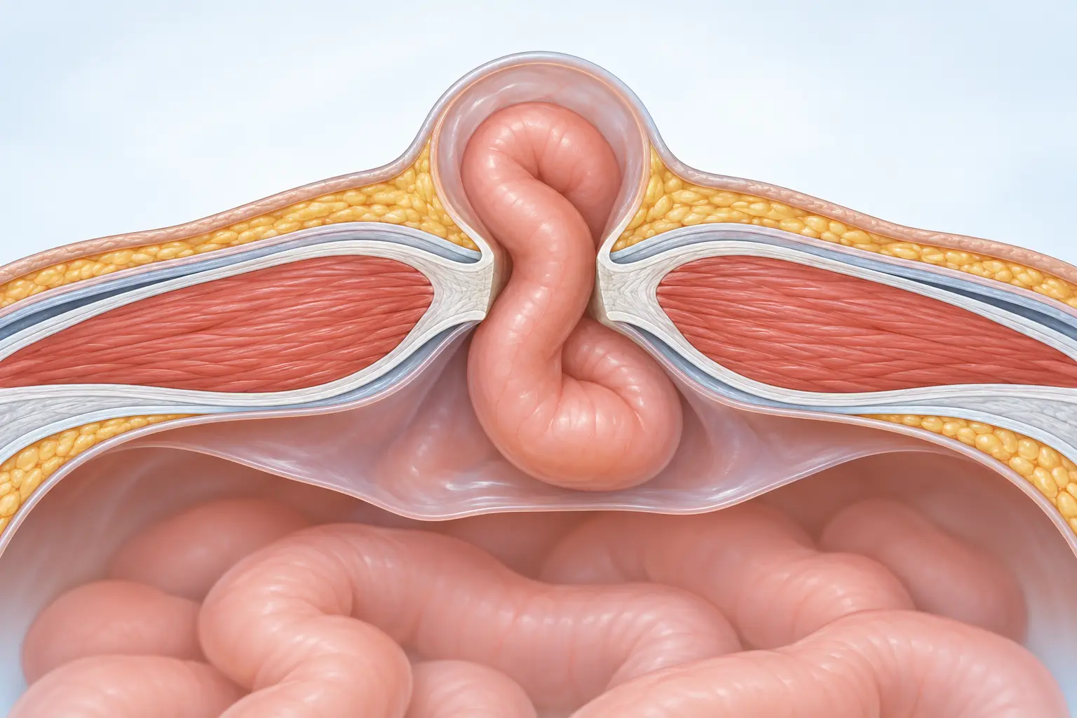
Congenital or acquired weak areas, particularly in the groin canal, umbilicus or abdominal midline.
A surgical scar may permanently weaken the fascia and become the site of an incisional hernia.
Chronic cough, constipation, obesity, smoking, diabetes and impaired wound healing can contribute to development or progression.
TYPES OF HERNIA
Location shapes the strategy.
Hernias are not all treated in the same way. Location, size, previous operations and abdominal wall quality determine the approach, reconstruction and mesh plane.
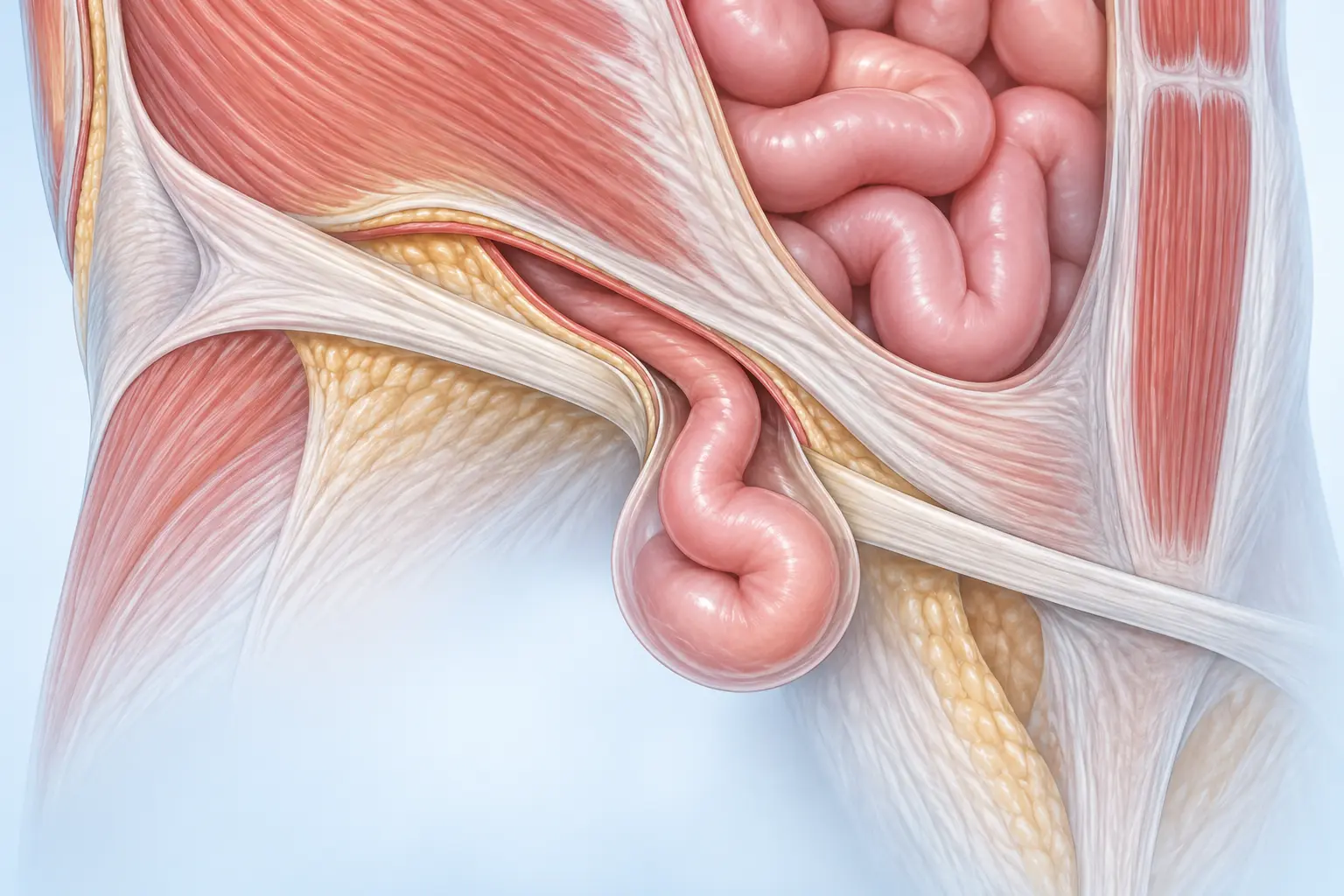
Inguinal hernia
The hernia sac protrudes in the region of the inguinal canal. Direct and indirect groin hernias are distinguished; in women, femoral hernia requires particular consideration.
Umbilical & ventral hernia
Primary ventral hernias arise without previous surgery, commonly at the umbilicus or above it in the midline.
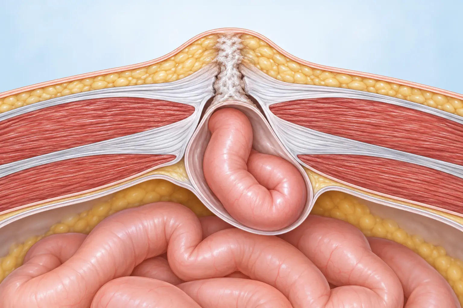
Incisional hernia
This develops at the site of previous abdominal surgery. For planning more complex cases, the EHS recommends cross-sectional imaging, usually CT or alternatively MRI.
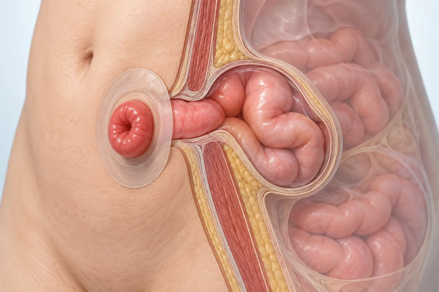
Parastomal hernia
The defect develops beside an intestinal stoma. Treatment and possible reconstruction must consider stoma function, symptoms and recurrence risk together.
When is urgent assessment needed?
Sudden severe or increasing pain, a firm irreducible bulge, nausea or vomiting, abdominal distension, inability to pass stool or wind, fever or reddened skin over the hernia may indicate incarceration or bowel involvement and require urgent assessment.
DIAGNOSIS
From examination to a reliable plan.
Many hernias can be identified clinically while standing and straining. Imaging supplements examination when the diagnosis is uncertain or the abdominal wall is complex.
Symptoms, progression, reducibility, previous operations and occupational demands.
Location, size, skin, stoma and abdominal wall function while lying and standing.
Ultrasound for selected questions; CT or MRI for uncertain, large, recurrent or complex incisional hernias.
Weight, smoking, diabetes, medication, lung function and individual operative risks.
TREATMENT OPTIONS
The least access necessary. The reconstruction required.
The goal is not simply to close an opening, but to achieve durable functional reconstruction. The procedure is tailored to the hernia and the patient, rather than chosen from technique preference alone.
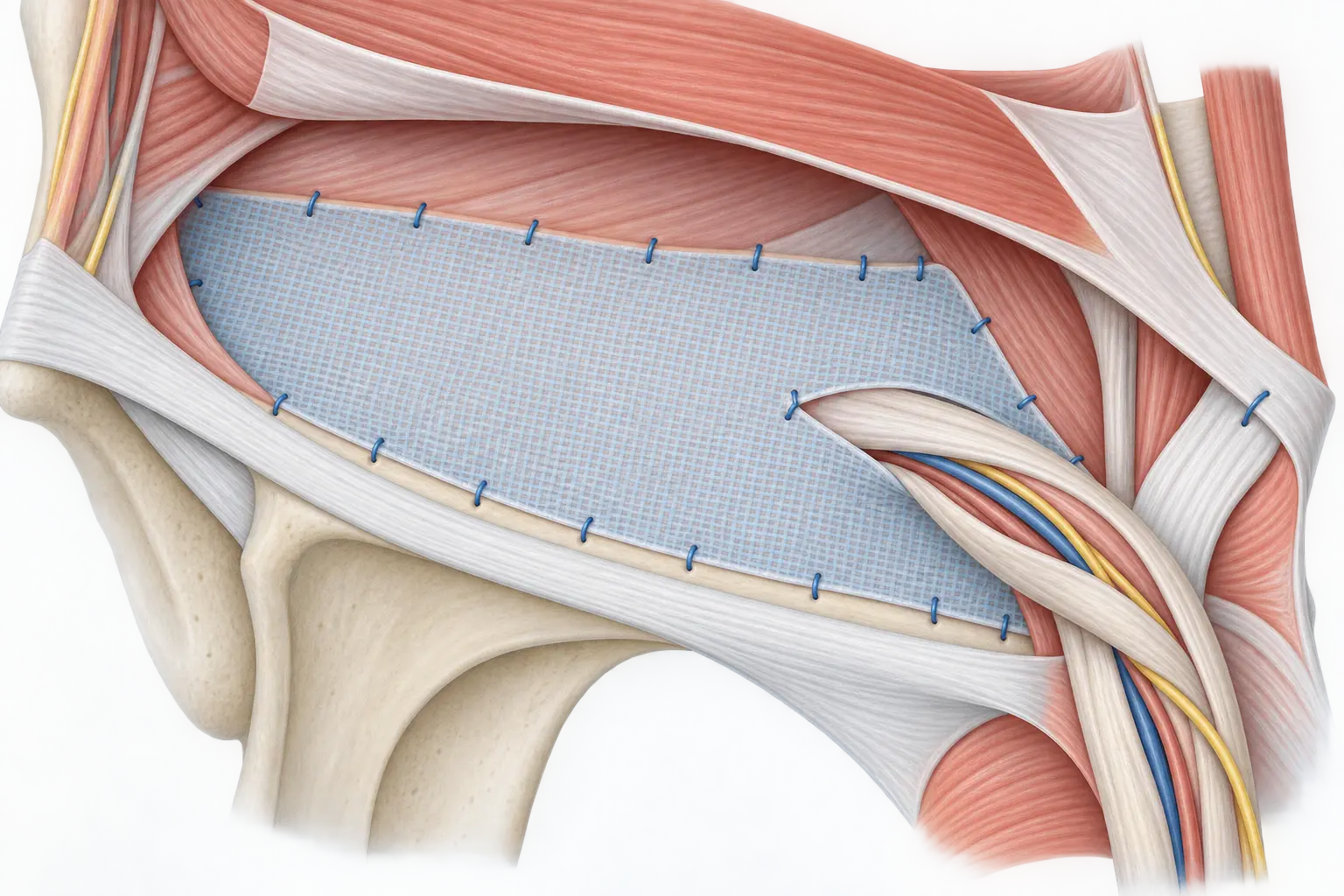
OPEN
Lichtenstein
An established open mesh repair of the groin. It may be appropriate for individual anaesthetic planning, certain previous operations or when a laparo-endoscopic approach is unsuitable.
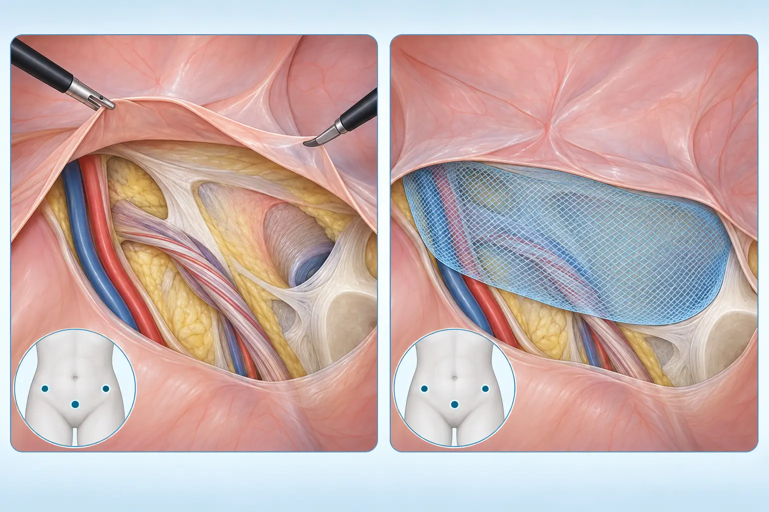
MINIMALLY INVASIVE
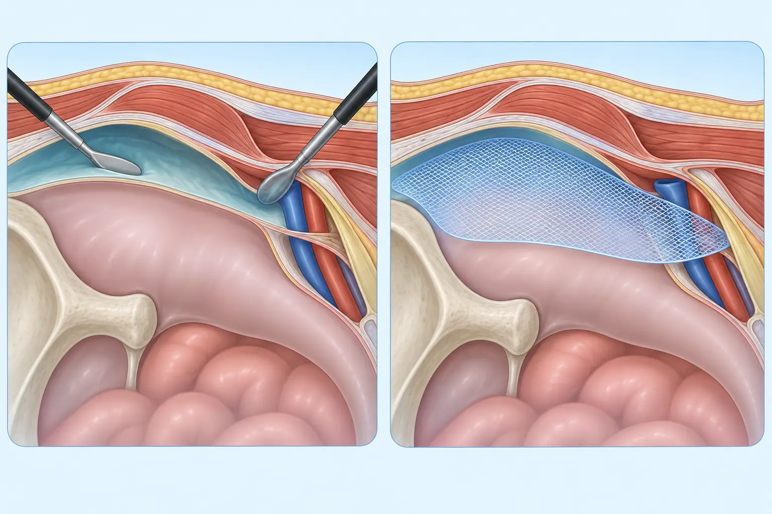
TAPP
In transabdominal preperitoneal repair, the abdominal cavity is inspected laparoscopically, the peritoneum is opened and a broad mesh is placed in the preperitoneal plane before the peritoneum is closed again.
EXTRAPERITONEAL
TEP
In total extraperitoneal repair, dissection takes place entirely outside the peritoneal cavity. The mesh covers the full myopectineal orifice within the preperitoneal plane.
TAPP or TEP?
Both are established laparo-endoscopic mesh techniques. They differ mainly in access rather than in their fundamental objective. The HerniaSurge guideline emphasises tailored procedure choice and surgeon expertise. Minimally invasive repair can be advantageous particularly for bilateral hernias, selected recurrences after an anterior repair and suitable primary hernias. Previous operations, hernia size, sex, general health and anaesthetic risk are considered.
ABDOMINAL WALL RECONSTRUCTION
Close the fascia. Place mesh in a protected plane.
For midline incisional hernias, the EHS recommends mesh-based reconstruction, preferably with retromuscular mesh placement. The fascial defect should be closed where possible and mesh bridging avoided.
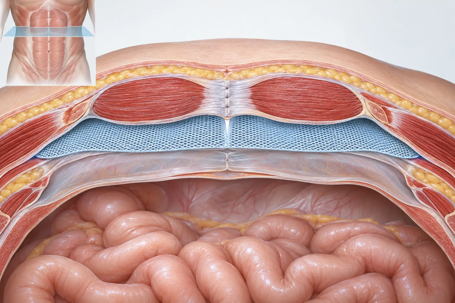
Sublay / retromuscular
The mesh lies behind the rectus muscles and outside the free peritoneal cavity. This protected plane allows broad reinforcement of the reconstructed fascia.
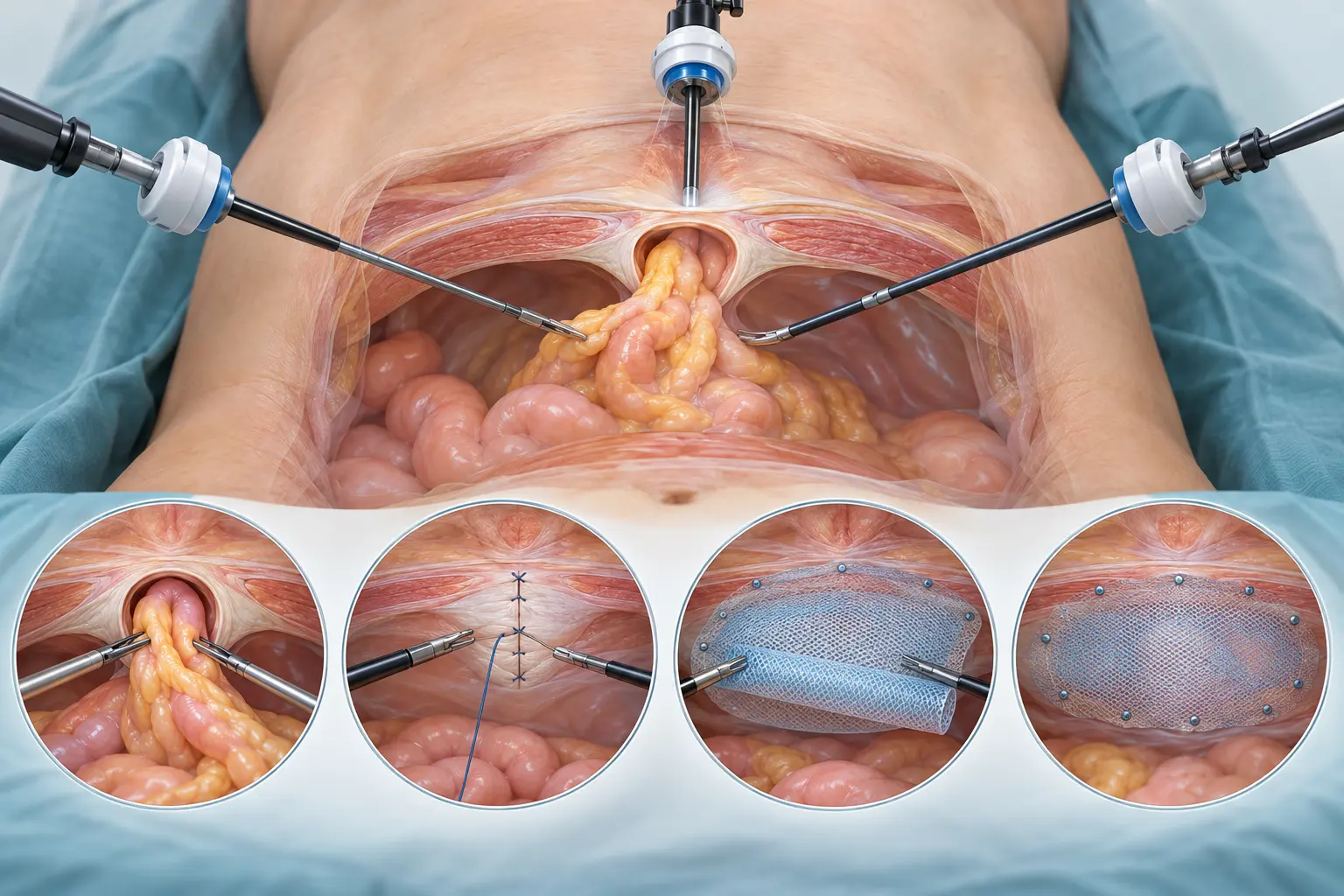
Minimally invasive reconstruction
Techniques such as eTEP or MILOS can reconstruct suitable abdominal walls through smaller access routes and allow extraperitoneal mesh placement.
Complex incisional hernia
Large defects, recurrences, multiple previous operations or loss of domain require detailed CT planning and, when necessary, advanced reconstructive techniques.
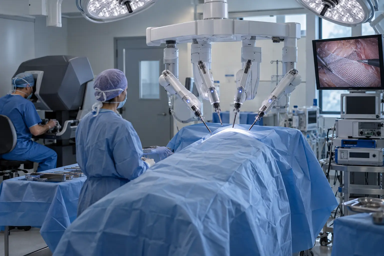
ROBOT-ASSISTED SURGERY
Precise reconstruction through small access points
Robotics is a surgical platform. It can facilitate controlled dissection within the abdominal wall, intracorporeal fascial closure and broad retromuscular mesh placement. Instrument articulation and stable three-dimensional vision may be helpful in more complex minimally invasive reconstructions.
- small access points in suitable hernias
- precise suturing and reconstructive steps
- potential for extraperitoneal mesh placement
- not automatic: benefit depends on anatomy and expertise
Putting evidence in context: the 2023 EHS guideline considers open, laparoscopic and robotic approaches potentially appropriate when matched to patient and hernia characteristics and supported by relevant expertise. Comparative long-term evidence is currently insufficient to claim general superiority of robotics.
AFTER SURGERY
Mobilise safely, increase activity gradually.
The pathway depends on access, hernia size and the extent of reconstruction. Personal discharge and activity advice takes precedence over general timelines.
The first days
- early mobilisation and regular short walks
- pain medication according to the agreed plan
- maintain hydration and avoid constipation
- keep wounds clean and follow dressing advice
Activity & work
- increase normal activity according to comfort
- drive only when reactions are safe and medication is not impairing
- obtain individual advice for sport, heavy lifting and physical work
- attend follow-up and use support garments if advised
What may commonly occur
- wound discomfort, tightness or temporary swelling
- bruising or fluid collection in the former hernia space
- temporary tiredness and reduced stamina
When to seek advice
- pain that increases rather than improves
- fever, chills, marked redness or wound discharge
- persistent vomiting, abdominal distension or inability to pass stool or wind
- shortness of breath, chest pain or one-sided leg swelling
Informed by European Hernia Society guidelines
This page summarises key recommendations in patient-friendly language. Guidelines support decision-making but do not replace individual assessment of anatomy, symptoms, risks and treatment goals.
HerniaSurge / EHS · Groin hernia management update · 2023 ↗EHS · Midline incisional hernia guidelines · 2023 ↗EHS / AHS · Umbilical and epigastric hernias · 2020 ↗EHS · Parastomal hernia prevention and treatment · 2018 ↗
Medical information: This content is intended for general orientation and does not replace personal medical examination, diagnosis or individual treatment advice. Acute warning signs require urgent medical assessment.
PROCTOLOGY & PELVIC FLOOR SURGERY
Proctology & Pelvic Floor Surgery
Modern diagnostics and individual treatment of conditions affecting the anal canal and rectum—with particular attention to function, continence and quality of life.
Individual treatment. Preserving function.
Proctology deals with conditions of the anal canal, rectum and the surrounding skin and soft tissues. Common conditions include haemorrhoidal disease, anal fissures, anal abscesses and anal fistulas. A pilonidal sinus—often called a pilonidal fistula—is also treated surgically, but is anatomically distinct from an anal fistula.
Many proctological conditions can be treated very effectively. In addition to established conservative and surgical approaches, minimally invasive techniques that preserve sphincter and healthy tissue are available. The aim is not simply to use the newest method, but to select the appropriate treatment based on symptoms, anatomy and stage of disease.
RECOGNISING SYMPTOMS
Typical symptoms
Proctological conditions can present in very different ways. A focused examination helps identify the cause reliably.
- Bright red blood during or after bowel movements
- Pain, burning or pressure in the anal area
- Itching, skin irritation or recurrent moisture
- Palpable swelling or lumps
- Mucous or purulent discharge
- Foreign-body sensation or tissue prolapse from the anal canal
- Difficulty with anal hygiene
- Recurrent inflammation or abscesses
- Changed bowel habits or difficulty with evacuation
These symptoms are not automatically caused by haemorrhoids. Rectal bleeding in particular should be medically assessed, as other bowel conditions can cause similar symptoms.
STEP BY STEP
Careful diagnostics
Assessment begins with a detailed discussion of symptoms, their duration, bowel habits, previous operations, medication and accompanying conditions, followed by a focused examination.
Inspection
Assessment of the outer anal region for swelling, inflammation, fistula openings, fissures, skin changes or prolapse.
Rectal examination
Careful digital examination of the anal canal and rectum when appropriate and tolerable.
Anoscopy / proctoscopy
Examination of the inner anal canal and lower rectum, particularly for assessing internal haemorrhoids.
Endosonography / MRI
For complex or recurrent fistulas to visualise the tract, branches and relationship to the sphincter.
Colonoscopy
May be required depending on age, bleeding pattern, family history, screening status and additional symptoms.
CONDITIONS & TREATMENT
Treatment tailored to the findings
Conservative, surgical and modern minimally invasive procedures are carefully balanced according to individual anatomy and stage of disease.
01 · CONDITION
Haemorrhoidal disease
What are haemorrhoids?
Haemorrhoids are normal, highly vascular cushions in the anal canal that support fine continence together with the sphincter. Haemorrhoidal disease occurs only when they enlarge or descend and cause symptoms.
Typical symptoms include bright red bleeding, itching, moisture, mucous discharge, pressure or a foreign-body sensation and tissue protruding during bowel movements.
- Constipation and straining
- Prolonged time on the toilet
- Frequent stools or chronic diarrhoea
- Pregnancy and increased pelvic pressure
Goligher classification
| Grade | Description |
|---|---|
| Grad I | Enlarged haemorrhoids without external prolapse. |
| Grad II | Prolapse during straining or bowel movements, with spontaneous reduction. |
| Grad III | Prolapse that must be manually reduced. |
| Grad IV | Permanently prolapsed tissue that cannot be reduced, or does not remain reduced. |
Conservative and outpatient treatment
In early disease, the priority is soft, formed stool without straining: a fibre-rich diet, adequate fluid intake, physical activity and treatment of constipation or chronic diarrhoea. Ointments or suppositories may provide short-term relief but do not correct the anatomical cause of advanced disease.
Rubber-band ligation places a small band over internal haemorrhoidal tissue so that it regresses. It is established particularly for symptomatic grade I and II disease and selected grade III cases. Sclerotherapy or infrared coagulation may also be considered depending on the findings.
Surgical procedures and laser treatment
Marked prolapse, larger external components or advanced disease may require haemorrhoidectomy. Targeted removal of enlarged nodules is highly effective and an established standard for advanced findings. Haemorrhoidal artery ligation closes supplying vessels and may be combined with mucopexy for mucosal prolapse; potentially less postoperative pain must be weighed against a higher recurrence risk depending on the initial findings.
Laser haemorrhoidoplasty (LHP) introduces a fine laser fibre into the enlarged cushion. Controlled energy coagulates and reduces the tissue. In selected grade II and III disease, studies show advantages in early pain, blood loss and recovery. Long-term evidence and recurrence outcomes are less consistent; LHP is a selective option, not inherently superior.
Prevention
Complete prevention is not always possible. Good long-term stool regulation, a fibre-rich diet, adequate fluid intake, regular activity, short toilet visits and avoiding straining can reduce symptoms and renewed stress.
02 · CONDITION
Perianal / anal abscess
Acute purulent inflammation
An anal abscess is an acute purulent inflammation around the anal canal and surrounding soft tissues. An abscess and a cryptoglandular anal fistula may represent different phases of the same process: the abscess is the acute phase, while a fistula tract may persist afterwards.
The most common cause is infection of small glands in the anal canal. Recurrent or atypical abscesses should be investigated for a fistula or another underlying condition.
- Subanodermal
- Intersphincteric
- Ischioanal / ischiorectal
- Supralevator or horseshoe abscess
Treatment and follow-up
A fully developed anal abscess generally cannot be cured with ointments or antibiotics alone. The key treatment is prompt surgical opening and adequate drainage. Antibiotics may be added for certain risks or systemic signs of infection, but do not replace drainage.
If a fistula is identified at the same time, further treatment depends on its course and sphincter involvement. Preserving sphincter function and continence is especially important. No reliable primary prevention is known; recurrent abscesses should prompt a targeted search for a fistula or underlying disease.
03 · CONDITION
Anal fistula
A tract between the anal canal and skin
An anal fistula is an abnormal tract connecting the anal canal or rectum with the skin around the anus. Many cryptoglandular fistulas develop after infection of anal glands or an anal abscess. Typical symptoms are recurrent pain, moisture, purulent discharge, itching or repeated abscesses.
- Intersphincteric fistula
- Transsphincteric fistula
- Suprasphincteric fistula
- Extrasphincteric fistula
Causes, classification and basic treatment
In addition to cryptoglandular fistulas, causes may include Crohn’s disease, previous surgery, injury or other conditions. Crohn-associated fistulas are often managed through interdisciplinary care. For surgical planning, the relationship to the sphincter is decisive: the more sphincter tissue involved, the more important a sphincter-preserving approach becomes.
A persistent cryptoglandular fistula usually does not heal reliably with medication alone. Acute infection may first require drainage or a seton. Superficial, low fistulas can be treated very effectively by fistulotomy provided there is no unacceptable continence risk. Higher or complex fistulas may be treated with advancement flap, LIFT, selected seton strategies, TROPIS, or endoscopic and laser-based procedures.
VAAFT · Video-Assisted Anal Fistula Treatment
VAAFT is a video-assisted, sphincter-preserving procedure. A thin fistuloscope is introduced through the external opening, allowing direct visualisation, irrigation and removal or coagulation of inflammatory tissue. Side branches can be identified and the internal opening treated or closed according to the findings.
Its advantage is direct endoscopic visualisation while largely preserving the sphincter. Evidence remains heterogeneous. VAAFT is a selective option, not a universal standard for every anal fistula.
FiLaC · Fistula-tract Laser Closure
FiLaC introduces a radially emitting laser fibre into the fistula tract. During controlled withdrawal, the tract wall is coagulated and contracted from within without dividing the sphincter. Healing rates vary and high-quality long-term data remain limited. FiLaC is therefore selected only after careful assessment of fistula anatomy.
Prevention and follow-up
No reliable primary prevention is known for cryptoglandular fistulas. Important measures include early treatment of an anal abscess, investigation of recurrent abscesses, treatment of underlying disease and consistent follow-up after surgery.
04 · CONDITION
Anal fissure
A tear in the delicate anal canal lining
Typical symptoms are severe cutting or burning pain during bowel movements that may persist for minutes to hours. A small amount of bright red blood is common. Hard stool, constipation, straining or repeated diarrhoea may contribute.
In chronic fissures, increased internal sphincter tension may impair local blood flow and healing. Atypical or multiple fissures should prompt assessment for underlying disease.
Conservative treatment
The priority is consistently soft stool through fibre, fluids, avoidance of straining and medication for stool regulation when needed. Warm sitz baths may relieve symptoms. In chronic fissures, topical nitrates or calcium-channel blockers such as diltiazem or nifedipine may reduce internal sphincter tension.
Botulinum toxin and surgery
Botulinum toxin injection can temporarily reduce muscle tension and support blood flow and healing, particularly after insufficient response to topical therapy. Lateral internal sphincterotomy is highly effective for chronic treatment-resistant fissures. Because sphincter surgery may affect continence, careful patient selection and alternative procedures where appropriate are essential.
Prevention
Normal, soft stool without straining is key. Adequate fibre and fluids, together with early treatment of constipation or persistent diarrhoea, can help prevent renewed mechanical injury.
05 · CONDITION
Pilonidal sinus
Pilonidal sinus—not the same as an anal fistula
A pilonidal sinus is an inflammatory condition of the skin and subcutaneous tissue in the cleft above the coccyx. Mechanical forces may drive hairs into the skin and cause chronic inflammation, small openings and branching sinus tracts.
- Asymptomatic disease
- Acute abscess-forming disease
- Chronic symptomatic disease
Selecting treatment
An asymptomatic finding does not require prophylactic surgery. An acute abscess requires surgical drainage, which should not be delayed by antibiotics. In chronic symptomatic disease, treatment depends on extent, number of openings, previous operations and individual factors.
Limited disease may be suitable for pit picking, sinusectomy, endoscopic treatment or laser ablation. More extensive or recurrent disease may be better managed in the long term with larger off-midline procedures.
EPSiT · Endoscopic Pilonidal Sinus Treatment
EPSiT introduces a thin endoscope through a small opening into the sinus system. Under direct vision, the tract and branches are assessed, hair and foreign material are removed, and inflammatory tissue is cleared or coagulated. The aim is small access points and limited tissue damage. Long-term evidence remains limited, and patient selection is crucial.
SiLaC · Laser ablation
SiLaC involves careful cleaning of the tract system followed by internal coagulation using a radially emitting laser fibre. Small skin openings and a limited wound area are potential advantages. Long-term comparative data remain limited, and laser treatment does not inherently replace larger operations in extensive or recurrent disease.
Prevention
No single preventive measure has proven universally effective. Good local hygiene, reduction of modifiable risk factors and early assessment of new symptoms are reasonable. Routine hair removal is not required for everyone and should be discussed individually.
TECHNOLOGY WITH JUDGEMENT
Modern minimally invasive proctology
Modern technologies broaden the treatment spectrum. They often aim to preserve the sphincter and healthy tissue while reducing postoperative burden.
| Procedure | Condition | Principle | Clinical context |
|---|---|---|---|
| Laser Hemorrhoidoplasty | Haemorrhoids | Laser coagulation and volume reduction | Minimally invasive for selected findings; long-term outcomes and recurrence risk must be considered. |
| VAAFT | Anal fistula | Endoscopic visualisation, cleaning and treatment | Sphincter-preserving; long-term evidence remains limited. |
| FiLaC | Anal fistula | Radial laser coagulation of the fistula tract | Sphincter-preserving; heterogeneous results make patient selection important. |
| EPSiT | Sinus pilonidalis | Endoscopic cleaning under direct vision | Small access points; an option for selected findings. |
| SiLaC | Sinus pilonidalis | Laser ablation of the cleaned sinus tract | Minimally invasive; long-term comparative evidence remains limited. |
IMPORTANT PRINCIPLE
Minimally invasive does not automatically mean superior.
The choice of procedure is guided by anatomy, stage of disease, sphincter involvement, recurrence risk, accompanying conditions and individual needs. The aim is not only to treat the disease, but to preserve function, continence and quality of life as effectively as possible. Benefits, limitations, risks and alternatives are discussed carefully before treatment.
SCIENTIFIC BASIS
Guideline-informed information
This patient information is informed by current German S3 guidelines and recommendations from international professional societies. Procedures with limited or heterogeneous long-term evidence are deliberately presented as selective options.
- S3 Guideline: Cryptoglandular Anal Fistulas · Version 3.0/2026 · AWMF 088-003register.awmf.org
- S3 Guideline: Anal Abscess · Version 3.0/2026 · AWMF 088-005register.awmf.org
- S3 Guideline: Pilonidal Sinus · Version 3.0/2026 · AWMF 081-009register.awmf.org
- ASCRS Clinical Practice Guidelines · Hemorrhoids (2024) & Anal Fissures (2023)fascrs.org
- Current systematic reviews of laser haemorrhoidoplastyPubMed
ROBOTIC AND MINIMALLY INVASIVE SURGERY
Progress that
enables precision.
Robotic & Minimally Invasive Surgery
With the da Vinci surgical system, I use advanced technology to plan minimally invasive surgery precisely and individually. Its suitability is always assessed according to the condition and the patient’s personal situation.
My experience in robotic surgery combines extensive operative expertise with the development and advancement of modern robotic surgery programs. A particular focus is complex colorectal surgery, where precise dissection, controlled suturing and the least traumatic approach possible are essential.
DA VINCI · ROBOTIC SURGERY
DA VINCI XI SURGICAL SYSTEM
Robotic Surgery in Motion
The da Vinci Xi Surgical System supports the surgeon during complex minimally invasive procedures through high-definition 3D visualization and highly articulated surgical instruments.
The surgeon's movements are precisely translated to the instruments. The surgeon remains in complete control of every movement and every step of the procedure at all times.
The video provides an insight into the technology and demonstrates the interaction between the surgeon console, robotic system and modern minimally invasive surgery.
The da Vinci system does not operate autonomously. Every movement of the instruments is controlled by the surgeon.
3D HD visionMagnified three-dimensional visualisation of anatomical structures.
Fine instrumentsHigh mobility and controlled movement transfer.
Minimally invasive accessSmall surgical access points when medically appropriate.
Individual planningEvery procedure is tailored to your situation.
YOUR PATH WITH DA VINCI
The treatment journey
PATIENT INFORMATION
How Robotic-Assisted Surgery Works
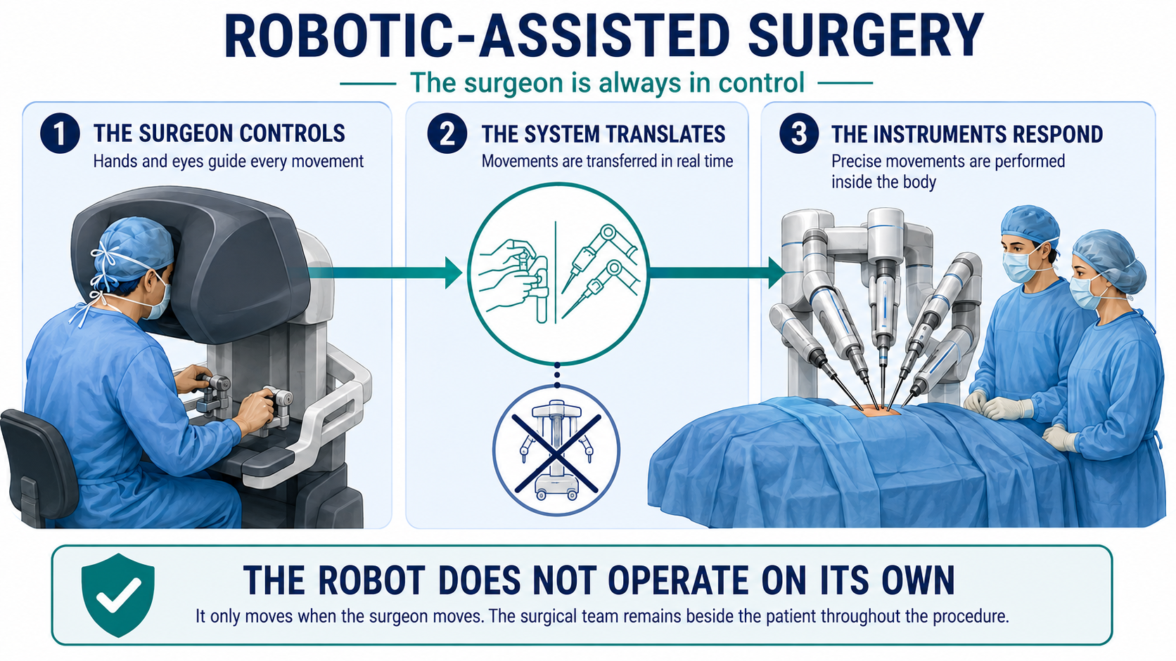
Robotic-assisted surgery is an advanced form of minimally invasive surgery. The system does not operate independently: a trained surgeon controls the camera and every surgical instrument throughout the procedure.
The surgeon is always in controlIf the surgeon stops moving, the instruments stop. The system does not make decisions or perform surgical steps on its own.
THE TECHNOLOGY
How the technology works
A robotic-assisted surgical system is a computer-controlled instrument platform consisting of a surgeon console, a patient-side unit with articulated arms, a high-definition camera and small surgical instruments. The surgeon and the operating-room team remain in the same room with the patient.
The surgeon controls the console
The surgeon views the surgical area in magnified three-dimensional detail and chooses every action: where to move, when to grasp tissue, when to cut and when to suture.
Movements are translated in real time
Hand movements are transferred immediately to the instruments. Motion scaling and tremor filtering can support precise work without replacing the surgeon’s knowledge, judgement or responsibility.
The instruments respond inside the body
A camera and fine articulated instruments are introduced through small access points. The surgeon uses the magnified view to identify anatomy and perform each operative step.
THE PROCEDURE
What happens during the operation?
Preparation & anaesthesia
General anaesthesia, safe positioning and standard safety checks are completed before the operation begins.
Small access points
The surgeon creates small incisions, inserts the ports and positions the robotic arms. Connecting the arms to the instruments is called docking.
Surgeon-controlled procedure
The surgeon performs the operation at the console. An assistant remains beside the patient, while the anaesthesia team continuously monitors the patient.
Completion & closure
The system is moved away and the small incisions are closed. A change to laparoscopy or open surgery may be made whenever the surgeon considers it safer.
POTENTIAL BENEFITS
Why robotic assistance may be helpful
Depending on the procedure and the individual patient, robotic assistance may offer enhanced visualisation, precise instrument control and improved access in confined spaces. Some patients may experience less postoperative pain, smaller scars or a faster recovery than after open surgery; these benefits vary and are not guaranteed.
Sources consulted in the patient information document: U.S. Food and Drug Administration, “Computer-Assisted Surgical Systems”; Kingston and Richmond NHS Foundation Trust, “Robot-assisted surgery”. This information does not replace personal medical advice.
Robotic & Minimally Invasive Visceral Surgery
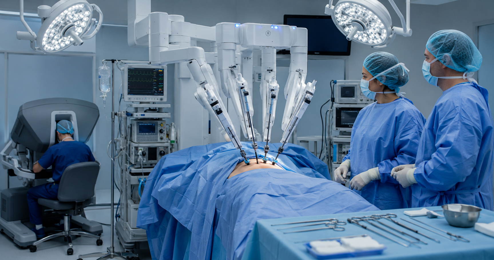
Modern visceral surgery combines advanced technology with proven clinical outcomes. Robotic and minimally invasive techniques represent the current standard in many complex abdominal procedures, offering significant benefits over traditional open surgery.
Smaller Incisions, Faster Recovery
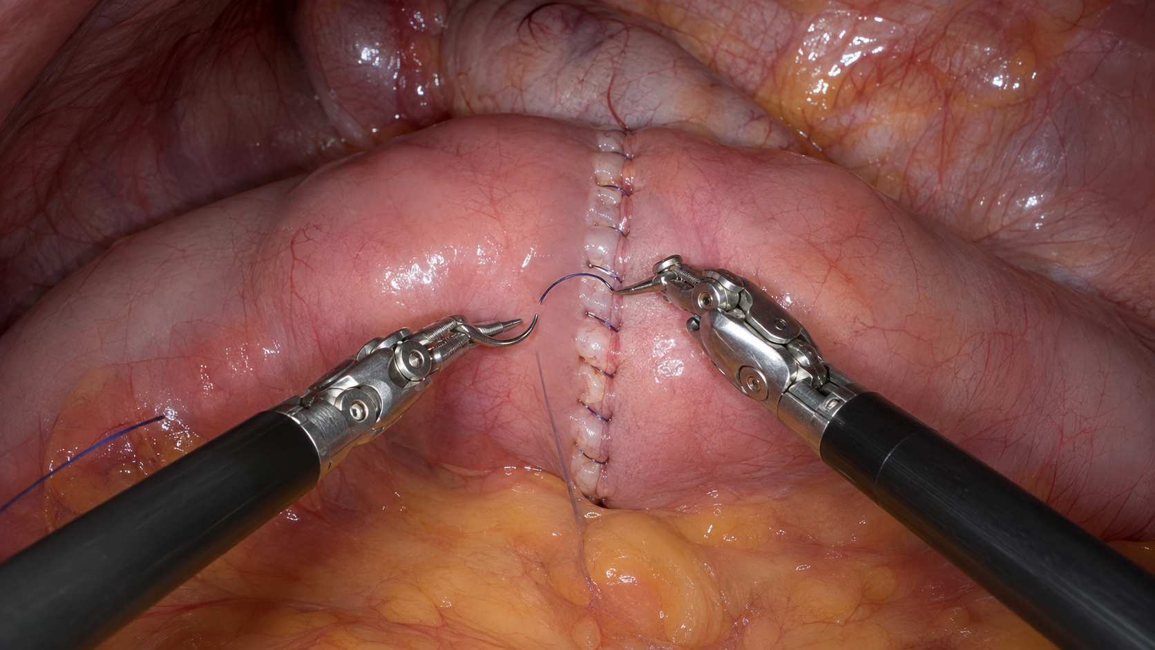
Minimally invasive surgery requires only small incisions (typically 5–15 mm for laparoscopy, similar or smaller for robotic assistance). This approach dramatically reduces tissue trauma, leading to shorter hospital stays, faster return to normal activities, and reduced postoperative pain. Studies consistently demonstrate that patients recover 2–3 times faster compared to open procedures.
Reduced Surgical Trauma & Complications
The minimally invasive approach decreases blood loss, reduces infection risk, and minimizes adhesion formation—a major cause of complications like bowel obstruction years after surgery. Prospective randomized trials, including the ROLARC study for colorectal cancer and the JALAPEÑO trial for gastric cancer, confirm that robotic and laparoscopic approaches achieve comparable oncologic outcomes to open surgery with fewer complications.
Enhanced Precision & Visualization
Robotic systems provide superior visualization with 3D, high-magnification imaging and instruments with greater dexterity and precision than the human hand. This is particularly valuable in complex dissections, anastomoses, and procedures requiring fine motor control. Laparoscopic instruments also offer magnified vision, enabling meticulous technique even in challenging anatomy.
Less Postoperative Pain & Fewer Scars
Smaller incisions mean minimal scar formation, reduced wound complications, and significantly lower pain levels postoperatively. Most patients require reduced analgesia requirements, improving quality of life immediately after surgery.
Equivalent Oncologic Outcomes
For cancer surgery (colorectal, gastric, and others), robotic and laparoscopic techniques achieve equivalent lymph node harvest, R0 resection rates, and long-term survival compared to open approaches. This has been validated in multiple randomized controlled trials and large prospective series, making these techniques the standard of care in high-volume centers.
Lower Impact on Pulmonary & Metabolic Function
The reduced inflammatory response and smaller incisions preserve core stability and respiratory function better than open surgery. Patients maintain better pulmonary function postoperatively and experience less systemic metabolic derangement, which is especially important in elderly or comorbid patients.
When Is Minimally Invasive Surgery Appropriate?
While most common visceral procedures (colorectal resections, gastrectomy, bariatric surgery, splenectomy, and others) can be performed safely with minimally invasive techniques, patient selection is crucial. Complex anatomy, emergency situations with hemodynamic instability, or extensive adhesions may warrant conversion to open surgery—and this is the mark of good surgical judgment, not failure.
Our Approach
We employ robotic and laparoscopic techniques as the standard approach for appropriate cases, with extensive training and proven outcomes. Every procedure is individualized based on patient factors, pathology, and operative findings. Our commitment is to provide the safest, most effective care with the smallest possible impact on your recovery.
For specific questions about your procedure and whether minimally invasive surgery is right for you, we encourage you to discuss this in detail during your consultation.
Evidence Base: This information is based on established guidelines from major surgical societies (SAGES, EAES), prospective randomized trials (ROLARC, JALAPEÑO), and current literature in visceral surgery. Individual outcomes vary based on patient factors and pathology.
Research & Publications
Academic work in robotic, colorectal, metabolic and minimally invasive surgery.
Robotic Surgery
Advancement of robotic surgical procedures and clinical treatment concepts.
Colorectal & Metabolic
Research projects in colorectal, bariatric and metabolic surgery.
Digital Surgery & AI
Perspectives for data analysis, quality improvement and intraoperative support.
Research & Publications
Academic work in robotic, colorectal, metabolic and minimally invasive surgery.
RESEARCH
Robotic Surgery
Advancement of robotic surgical procedures and clinical treatment concepts.
CLINICAL SCIENCE
Colorectal & Metabolic
Research projects in colorectal, bariatric and metabolic surgery.
INNOVATION
Digital Surgery & AI
Perspectives for data analysis, quality improvement and intraoperative support.
SCIENTIFIC WORK
15 Recent Publications
Selected from the latest PubMed records for Omar Thaher. Open any publication to read the complete entry on PubMed.
01
Timing of a laparoscopic cholecystectomy in acute moderately severe to severe biliary pancreatitis
PUBMED ↗ 02Thromboprophylaxis, pain and organization: How do expert centers manage PIPAC's perioperative care? An international survey
PUBMED ↗ 03An international multidisciplinary consensus statement on laparoscopic liver biopsy
PUBMED ↗ 04Esophageal Reflux Barrier Might be More than Just An Anatomical Definition: A Systematic Literature Review
PUBMED ↗ 05Gender-specific Differences in Preoperative Characteristics and Perioperative Outcomes of Patients Undergoing Robotic Resection Rectopexy With NOSE for Obstructive Defecation Syndrome
PUBMED ↗ 06Minimally invasive resection rectopexy as a treatment method for obstructive defecation (ODS): functional outcome in ODS; constipation and fecal incontinence
PUBMED ↗ 07Thromboembolic prophylaxis in bariatric and metabolic surgery: state-of-the-art according to the results of a nationwide registry study
PUBMED ↗ 08Burden of 375 diseases and injuries, risk-attributable burden of 88 risk factors, and healthy life expectancy in 204 countries and territories, including 660 subnational locations, 1990–2023
PUBMED ↗ 09Outcomes of Endoscopic Sleeve Gastroplasty: A Systematic Review
PUBMED ↗ 10Lipedema after Bariatric and Metabolic Surgery: A Scoping Review
PUBMED ↗ 11Vascular and Anatomical Challenges in Renal Transplant Surgery; What a Urologist Needs to know
PUBMED ↗ 12Predictive modeling for metastasis in oncology: current methods and future directions
PUBMED ↗ 13Short- and mid-term functional outcomes of STARR procedure in obstructed defecation syndrome
PUBMED ↗ 14Robot-assisted mesh rectosacropexy and sigmoid colon resection for obstructive defecation syndrome: a two-stage surgical approach
PUBMED ↗ 15Erratum: Burden of disease attributable to high body mass index: an analysis of data from the Global Burden of Disease Study 2021
PUBMED ↗Publication data verified via PubMed. Records are ordered by PubMed publication date, newest first.
Patient Information
Clear guidance before, during and after planned treatment.
Before Surgery
Important information about preparation, medication and admission.
Your Hospital Stay
Processes, ward rounds and the essential steps surrounding your operation.
After Discharge
Information on activity, nutrition, wound checks and follow-up appointments.
PATIENT INFORMATION
Patient Information for Surgery
Clear, evidence-informed guidance before, during and after planned surgery—with a particular focus on preparation, risk reduction and safe recovery.
KEY POINTS AT A GLANCE
How you can support your recovery
Many factors that influence the course of surgery and recovery can be improved before the procedure. These include stopping smoking, reducing alcohol, regular activity, adequate hydration, balanced nutrition and structured preparation with a medication review and checklists.
Modifiable risk factors should be identified as early as possible and, where feasible, reduced before planned surgery.

BEFORE SURGERY
Preparing well for your procedure
Good organisation, healthy habits and a complete medical overview help reduce avoidable risks.

ORGANISATION
Preparing for surgery
Good organisational and medical preparation helps prevent errors and makes the day of surgery run more smoothly. This includes the preoperative assessment, required laboratory tests, a review of your medication list and all documents needed for admission.
- Bring an up-to-date medication and allergy list
- Bring relevant reports, letters and identification
- Follow the fasting instructions from your care team exactly
- Arrange transport and an escort home if advised

RISK REDUCTION
Stopping smoking before surgery
Smoking increases the risk of pulmonary and cardiovascular complications, wound-healing problems and infection. Even a few weeks without nicotine can improve tissue oxygenation and healing.
Recommendation: stop as early as possible—ideally at least four weeks before surgery.

HEALTH HABITS
Reducing alcohol intake
Regular or high alcohol intake can increase perioperative risk. It may affect the immune system, liver function, blood clotting and the response to anaesthetic medicines.
Please be open about your actual intake. This allows the care team to assess risks safely and support you individually.

NUTRITION
Nutrition and malnutrition
Good nutritional status is particularly important before major surgery. Tell your care team if you have lost significant weight in recent months, have little appetite or can eat only very small amounts.
- Aim for adequate protein and energy intake
- Identify malnutrition early
- Use dietetic support or oral supplements if recommended

BLOOD TESTS
Identifying anaemia and iron deficiency
Preoperative anaemia is not uncommon and should be identified as early as possible before major surgery. Common causes include iron deficiency, chronic inflammation, vitamin deficiency and other underlying conditions.
Timely testing can identify the cause and, where possible, allow treatment before surgery.

MEDICATION SAFETY
Medicines before surgery
Your care team needs a complete medication list. Blood thinners, diabetes medicines, insulin, steroids, heart medicines, painkillers, herbal preparations and supplements are particularly important.
Never stop medicines on your own. Which medicines should be paused, adjusted or continued depends on the procedure and your individual risk and will be decided by your surgical or anaesthesia team.
IN HOSPITAL
Safety and early recovery
Effective pain management, early mobilisation and structured care are central parts of your treatment pathway.

TREATMENT PATHWAY
During your hospital stay
In hospital, the focus is on safety, effective pain relief, early mobilisation and structured care. As soon as medically appropriate, drinking, eating and movement are gradually resumed.
- Risk-adapted thrombosis prevention
- Multimodal pain management
- Early mobilisation
- Early oral fluids and food when medically appropriate
AFTER DISCHARGE
Continuing your recovery safely at home
Wound care, adequate hydration, healthy nutrition, gradual activity and follow-up appointments support your ongoing recovery.

RECOVERY
Aftercare and wound care
Recovery does not end when you leave hospital. Follow your personal discharge plan and increase activity gradually. Attend arranged wound checks and follow-up appointments.
- Care for the wound according to your individual instructions
- Drink enough and eat a balanced diet
- Increase activity gradually
- Attend scheduled follow-up appointments
Your personal discharge plan
Your care team will provide instructions on wound care, showering, physical activity, nutrition and medicines. These individual instructions take priority.
Warning signs—seek prompt assessment
- Increasing redness, swelling or pus-like discharge
- Worsening pain or persistent bleeding
- Fever, shortness of breath, marked weakness or circulatory problems
Guideline-informed patient information
The content is informed by established guidelines and studies on ERAS®, perioperative risk reduction, nutrition, anaemia management and infection prevention.
ERAS® – Enhanced Recovery After Surgery
ERAS combines structured preoperative preparation, modern anaesthesia and surgical techniques, multimodal pain management, early mobilisation and early nutrition in a standardised pathway.
Smoking cessation
The WHO describes benefits particularly when smoking is stopped around four weeks or more before surgery.
Nutrition and anaemia
ESPEN and international consensus recommendations support early identification and treatment of malnutrition and preoperative anaemia.
ERAS Society ↗ESAIC ↗WHO · Smoking & Surgery ↗WHO · Surgical Site Infection ↗Cochrane ↗PREHAB Trial ↗Anaemia Consensus ↗
Updated August 2026. This information is for general guidance and does not replace individual medical advice, surgical consent or anaesthetic assessment. For acute or severe symptoms, please contact your local emergency service or the nearest emergency department.
Your treatment begins with a personal consultation.
Personal surgical consultation · by individual appointment
CONTACT & APPOINTMENT
Contact & Appointment Request
For a personal surgical consultation, please submit your request through the form. Your enquiry will then be reviewed individually.
Request an appointment
Please complete the details below. Required fields are marked with *.
Your details are used exclusively to process your enquiry.